Genus Zonomyxa Nüsslin, 1882
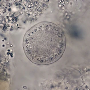
Diagnosis: Shell discoid in the resting stage, 140–160 µm; locomotive form pyriform or oblong, 220–250 µm long. Shell a chitinoid pellicle without a mucilaginous envelope; flexible and following the movements of the cell. Surface with small, temporary perforations through which thin plasma threads may emerge. Pseudopods single, clear, conical, arising from a slit‑like pseudostome. Endoplasm granular and violet‑tinted. Up to 32 vesicular nuclei reported. Crystalloid bodies 3–5 µm.
Feeding: herbivorous.
Ecology: Sphagnum mosses. Monospecific.
Zonomyxa violacea Nüsslin, 1882
Diagnosis: As for the genus. The description is almost identical to that of Amphizonella violacea. According to Meisterfeld (2006), Zonomyxa violacea can be distinguished from A. violacea by the following characters:
– a thinner membrane
– absence of an external mucus layer
– and, most significantly, the presence of numerous nuclei
Zonomyxa typically has four nuclei (Meisterfeld, 2006), although Penard (1906) reported up to 32. These nuclei (~20 µm) are smaller than the single large nucleus of Amphizonella violacea. I found an average of seven nuclei per cell. However, it remains uncertain whether Zonomyxa is truly distinct from Amphizonella.
Dimensions: My measurements: 360–450 µm; nuclei approx. 28 µm.
Ecology: in Sphagnum. Tirol, France.
Remarks: I have only found cells containing around seven spherical nuclei. Nuclei are difficult to detect in living specimens; I crushed the cells to force the nuclei out of the cell membrane. My specimens were collected from Sphagnum in Tirol, Austria, and I have also found this species in Sphagnum from the Vosges, France.


Compare the above specimen with the original drawing below made by Nüsslin (1884). This drawing shows much better the common shape of this organism as the one which is usually used in textbooks, a drawing made by Penard.

The drawing above was made by Penard (1906) and is usually used in textbooks, adding more confusion than clarity to the identity of this amoeboid. Penards drawing closely resembles the one of Nüsslin in the upper right corner of the drawing below, where it has the shape of a <. Remarkable are the thin ‘hairs’ around the cell. I’ve seen similar ‘hairs’ in specimens of A. violacea, where they were bacteria along wires of mucus.