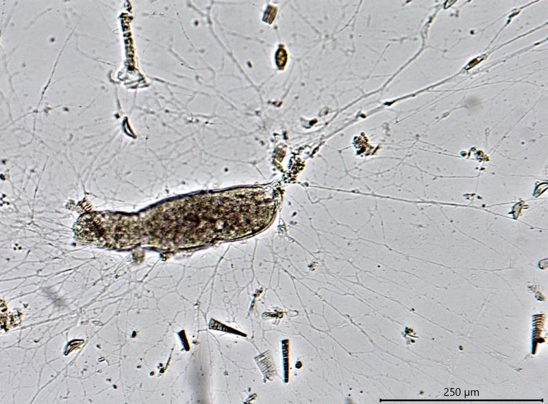
Lieberkuehnia spec., in Petri dish, sausage shaped
Lieberkuehnia sp. nov.
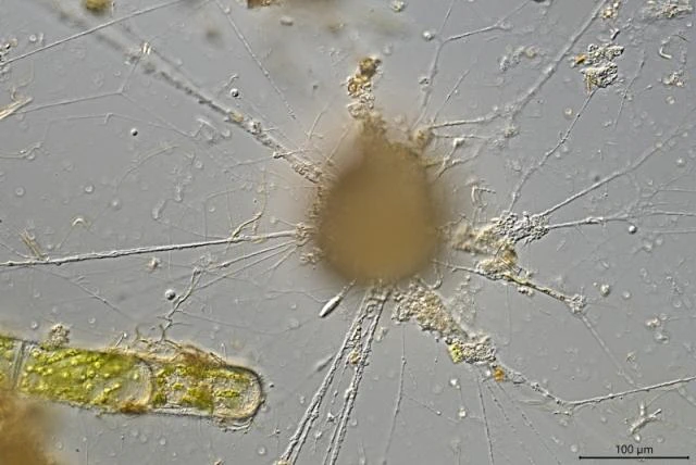
Diagnosis: Cell large, up to 340 µm, commonly sausage-shaped, with irregular outline and an organic-walled membranous test with a single aperture. Shell hyaline and colorless, flexible, smooth and delicate. From a long ribbon-shaped trunk long branching and anastomosing granuloreticulopodia radiate with continuous bi-directional streaming of granules. Cytoplasm with many small bi-refringent yellowish crystal-like bodies (ca. 1.5-2.3 µm long), food vacuoles and nuclei. Nuclei spherical, 7.9-9.5 µm (mean 8.6 µm; n=15) in diameter, each nucleus with some peripheral nucleoli.
Measurements. 157-340 µm (n=5)
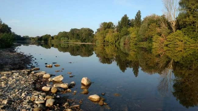
Ecology: Freshwater; in sediment of the shallow part of the river Gardon along camping Beau Rivage near Cardet, France (September 2019)
Remarks: I found six specimens in Petri dishes. In a living cell, transported to a slide and slightly compressed by the cover glass, the nuclei were light microscopically undetectable, but as the cell was compressed even more, cytoplasm burst out of the cell and the nuclei contained therein became immediately visible.
Cell with long peduncle
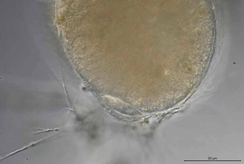
Peduncle in cross section, lens-shaped
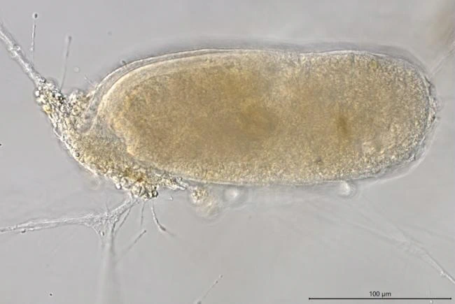
Large specimen
Specimen in wet mount, more or less spherical; granuloreticulopodia over 1 mm long.
The same specimen as above.
Specimen in a Petri dish. It had a kind of ‘tail’, probably a remnant of a division (see combined pictures below, cell A)
The same specimen as above. It was impossible to detect nuclei in this cell. Only when it was compressed by pressing the cover glass, plasm burst out of the cell and nuclei became clearly visible.
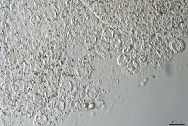
Nuclei, about 40 in the specimen above
Nuclei
The same specimen as above, but now with (yellow) crystals better visible.
Yellow bi-refringent crystal-like bodies
This is one of the two cells (B) on the photographs below.
This combination of four photographs show two cells of Lieberkuehnia sp. (A, B) which are connected by some thin long granuloreticulopodia. Probably both cells did divide and moved away from each other. The distance between both cells was almost 2 mm.
The Gardon river in France, home to this new Lieberkuehnia species. The sample location is between the two large stones in the water.