Omnivora mutabilis (Bailey, 1853) Dumack, Pundt and Bonkowski, 2019
Basionym: Pamphagus mutabilis Bailey, 1853
Synonym: Pseudodifflugia caudata Penard, 1910
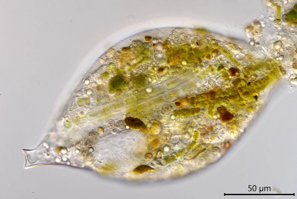
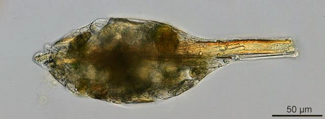
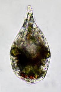
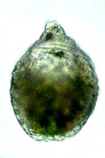
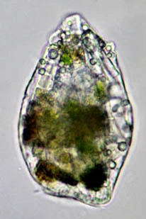
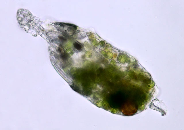
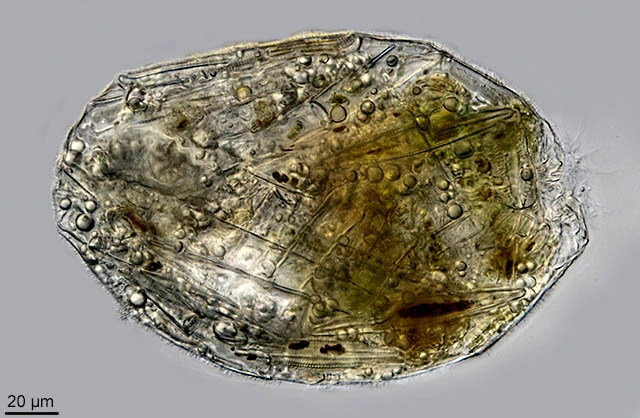
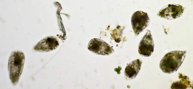
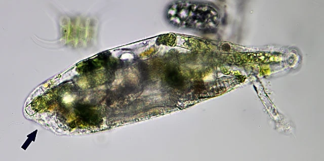
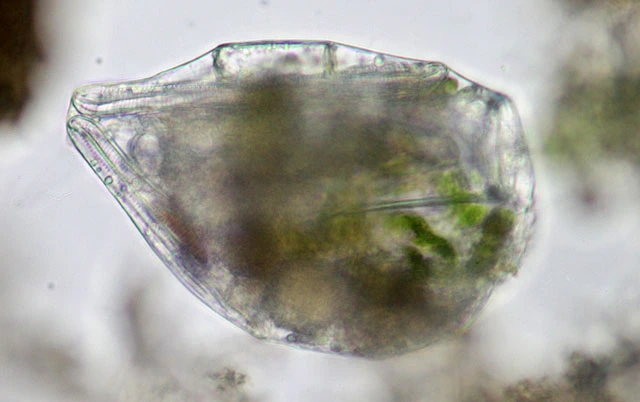
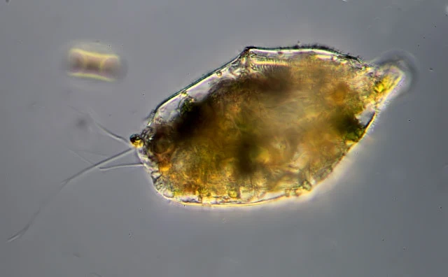
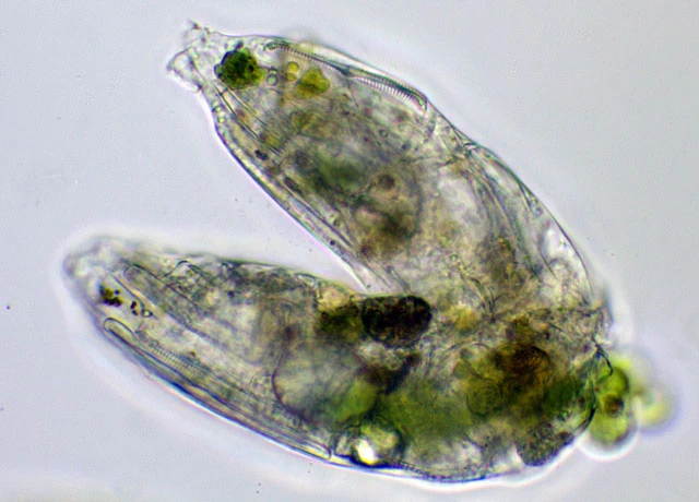
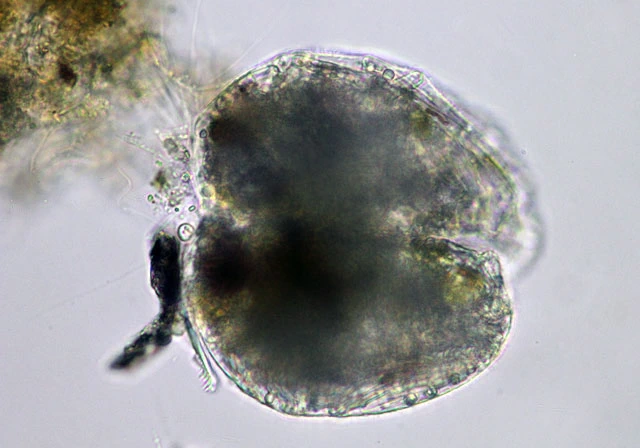
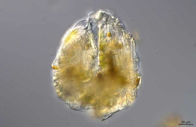
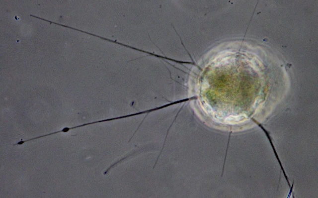
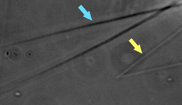
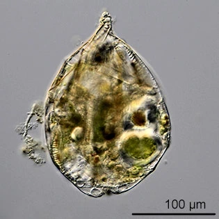
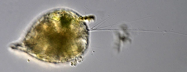
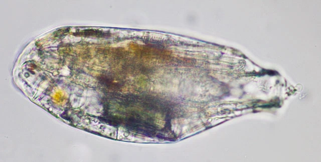
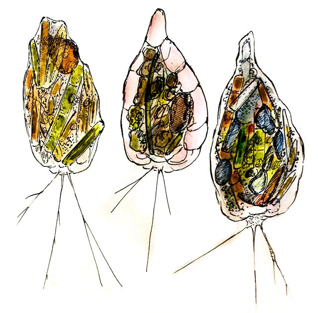
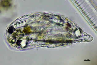
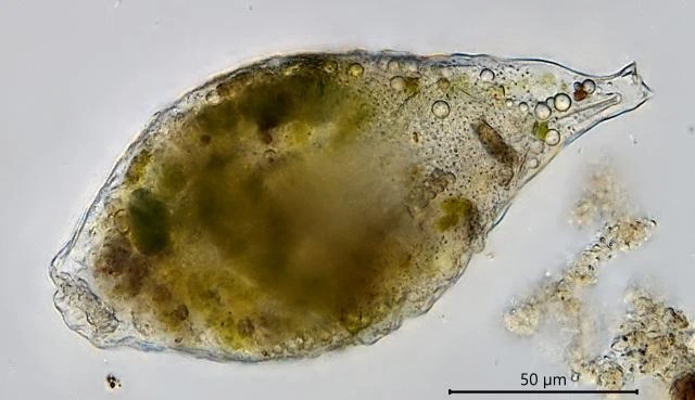
Diagnosis: test a highly flexible, colorless, hyaline membrane, following the size changes of the cytoplasm during feeding or starvation; shape usually pyriform or droplet shaped, with the pseudostome on the broad base; filopodia usually few, long and strait, branching. Large filopodia slightly taper, smaller filopodia are rod shaped, with parallel sides. Longitudinal division.
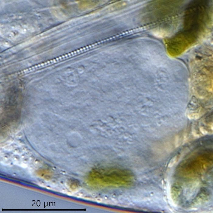
Dimensions: 103-150 µm long; my measurements: 133-264 µm; nucleus c. 33 µm.
Habitat: In different types of water, on sediments and between aquatic plants; easily overlooked because of the resemblance with a piece of debris. I’ve also found it in eutrophic water. Food: mostly different kind of algae and diatoms.
Remarks: the pseudostome is hard to observe; it is usually a small slit and often hidden between folds of the membrane.
Originally this curious organism has been described as Pamphagus mutabilis. The use of the name Pamphagus is not correct according to the International Rules, as pointed out by Wailes (1915).
Several authors has described a species under this name, but only few has apparently seen what Bailey described as follows: “If the reader will imagine a bag made of some soft extensible material so thin as to be transparent like glass, so soft as to yield readily by extension when subjected to internal pressure, and so small as to be microscopic; this bag filled with particles of sand, shells of diatoms, portions of algae or desmids, and with fragments of variously colored cotton, woolen, and linen fibers, will give a picture of the animal; to complete which it is only necessary to add a few loose strings to the bag, to represent the variable radiant processes which it possesses around the mouth.”
Penard (1910) described this amoeboid as Pseudodifflugia caudata.