
A. pedatus, drawing after Penard, 1903
Genus Actinosphaeridium Zacharias, 1893
Actinosphaeridium pedatus Zacharias, 1893
Syn.: Nucearia caulescens Penard, 1903
Diagnosis: Cell surrounded by a mucous layer. Filopodia with granules, sometimes branching. Cell stalked; stalk ends at the cell body. During cyst formation the cyst is covered with siliceous plates.
Dimensions: Cell body 16-20 µm, with mucous layer about 40 µm.
Ecology: Several specimens between wet moss in a small pond on an ecoduct near Crailoo, the Netherlands, February 2021, just one week after the ice had melted.
Remarks: I could not detect any mucous layer in my specimens. It was not visible in phase contrast, nor in DIC. The cells were surrounded by a thin membranous shell, which was hard to detect.

Actinosphaeridium pedatus
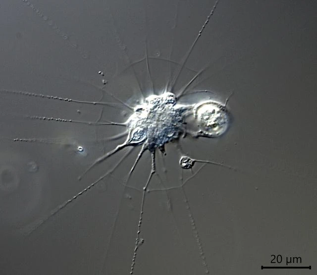
Habitus, branched pseudopodium

Cell with captured prey


“Foot” of the stalk, attached to the cover glass

Phase contrast