Astroperula parvipila Siemensma and Holzmann, 2023
Diagnosis: Multinucleate organic-walled monothalamous foraminifera. Cells spherical to pyriform bi-symmetrical with curved dorsal side and flatter ventral side. Shell wall flexible, colorless and smooth. Aperture terminal, oblique. Peduncle more or less trumpet-shaped, eccentrically located and varying in length, surrounded by a hyaline peduncular sheath. Radiating network of granuloreticulopodia. Cytoplasm with numerous yellowish crystalline birefringent crystalline rod-shaped bodies.
Ecology (Type locality): On submerged basalt rocks at the base of the dike along the northern part of Gooimeer, a lake in the Netherlands (52°18’18.5″N 5°18’50.1″E).
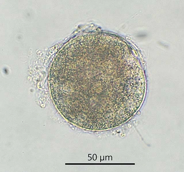
Description. The test is monothalamous and organic walled, hyaline and colorless, flexible, smooth and delicate. Diameter of the cell is 65 µm. The cytoplasm contains numerous yellowish birefringent rod-shaped crystalline inclusions. A nucleus or nuclei could not be detected, possibly obscured by the numerous inclusions.
Differential diagnosis. Astroperula parvipila can be distinguished from A. dumacki by its smaller size, about 65 µm vs. about 122 µm, with the restriction that this difference is only based on one specimen of each species. It can be distinguished from L. wageneri and C. lachmannii by the absence of yellow, birefringent rod-shaped crystalline inclusions in the latter two species. Based on observed morphological characters a clear distinction of A. parvipila and Velamentofex spp. is not possible.
The 18S barcoding sequence of A. parvipila contains 1141 nucleotides, the GC content is 41%.
Observed food. Diatoms.