Claparedellus lachmannii
Specimens of C. lachmannii has been found in the Netherlands, in a large lake called Gooimeer.
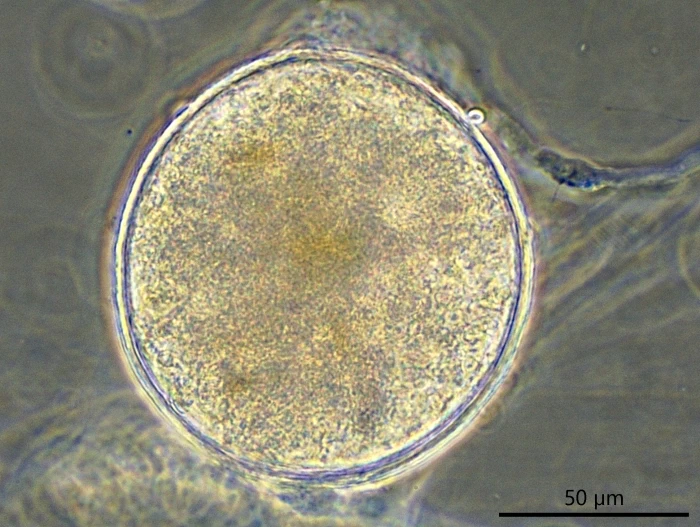
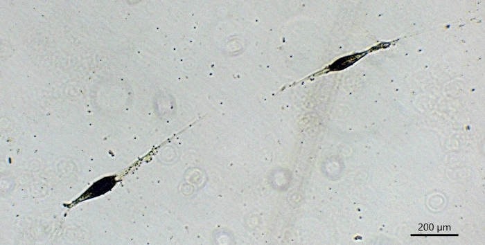
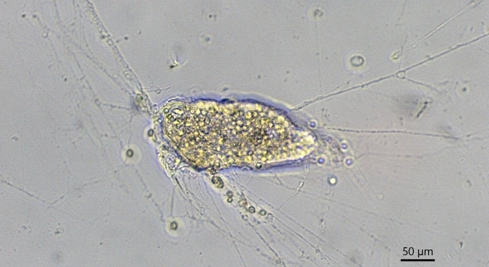
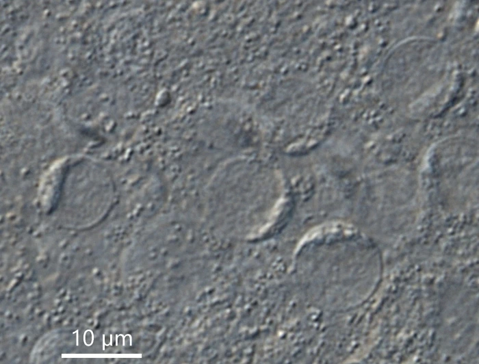
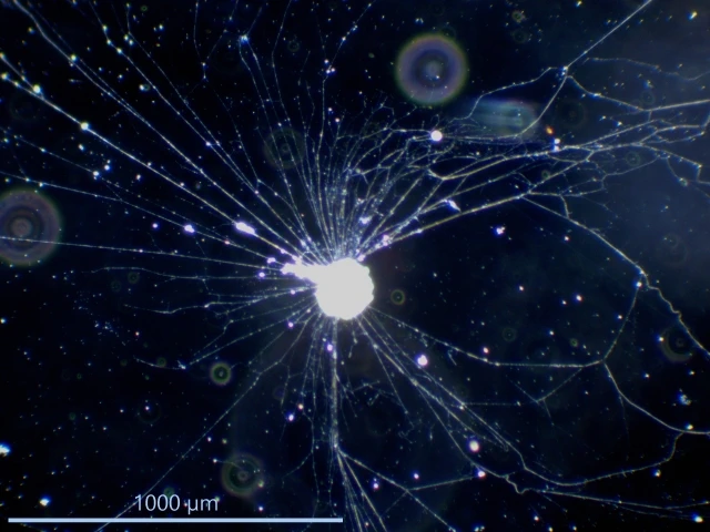
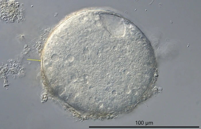
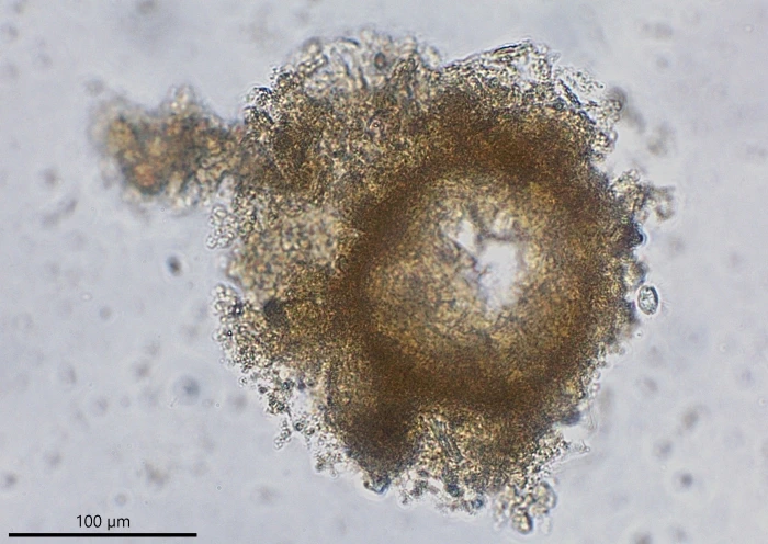
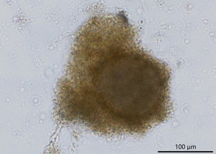
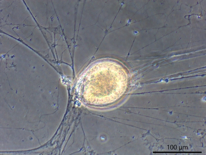
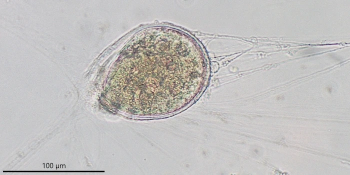
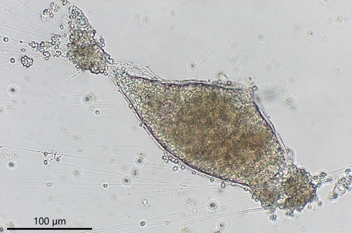
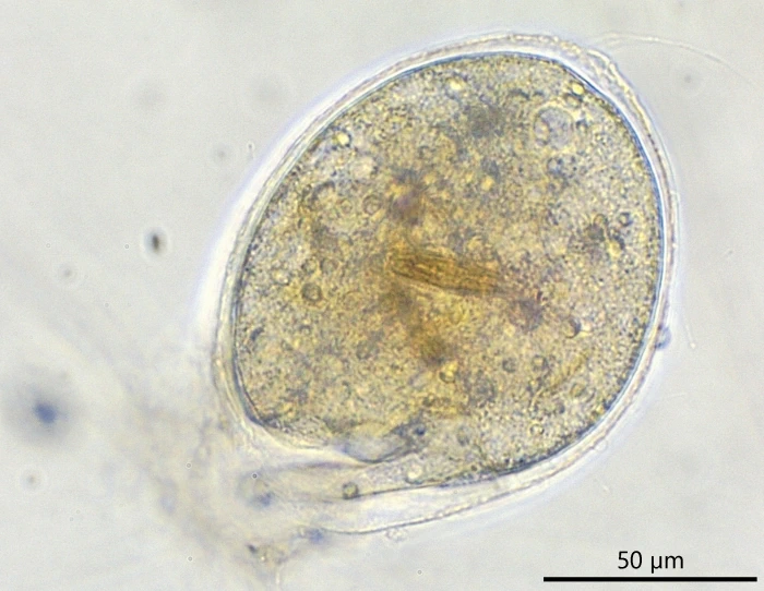
Description of the Gooimeer population. Stationary cells are spherical, locomotive cells are spherical to ovoid, sometimes elongated, fusiform or tubular, and sometimes wrinkled. The size of 15 cultured specimens that all derived from one cell, varies between 92-146 µm (mean 111 µm). A few long granuloreticulopodia radiate from the peduncle, forming one or two leading strands and one or more trailing strands with a length of 5,000 µm or more. The cytoplasm contains food vacuoles, some contractile vacuoles, and numerous colorless spherical granules, up to 0.8 µm in diameter. Forty-three nuclei were counted in one specimen, similar numbers were observed in other C. lachmannii. Each nucleus is spherical in shape with a disc-shaped nucleolus, which is close to the nuclear membrane. The nucleolus can take the shape of a crescent or spherical plate depending on its position. The nuclear plasma shows a granular structure when observed with a 100X oil immersion objective. The diameter of the nuclei, measured in three cells, is 6.0-7.5 µm (mean 6.5 µm, n=15), 6.1-7.8 µm (mean 7.0 µm, n=15) and 6.7-7.9 µm (mean 7.3 µm, n=15) respectively.
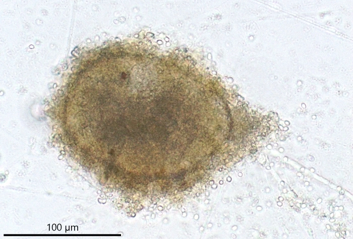
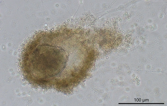
Stationary cells are occasionally covered with an accumulated layer of debris and excreted digested material. They can stay under this protective cover for several days. The cover is not a firm structure and could sometimes be removed when gently pipetting culture medium in its direction. It is left behind when stationary cells start to move again. Cells might gather together under a protective cover for reproduction as has been documented for Velamentofex dujardini and other Velamentofex species (Siemensma et al., 2021). No such observation has been made for C. lachmannii, although it cannot be excluded that the species uses its cover to reproduce underneath.
Observed reproduction. A part of the cell division could be observed for one specimen that was extremely elongated, measuring almost 300 µm in length. The hyaline part connecting the two daughter cells subsequently became longer and thinner, and after several hours both cells finally separated. During division, both cells moved like conjoined twins. As we could not determine the beginning of cell division, we do not know how long the whole process takes.
Observed food. Diatoms.