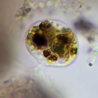
Frenzelina reniformis, at two diffferent levels
Frenzelina reniformis Penard, 1902
Diagnosis: Shell membranous, semi-hemispherical or hemispherical, usually with an open ventral face; very small mineral grains, loosely attached to the membrane; test in dorsal view usually elliptical; test not always present; plasmabody broad-elliptical or kidney-shaped; aperture flexible, commonly a short small tube; numerous fine filopodia, often in a bundle; nucleus vesicular with one central nucleolus.
Dimensions: 26-30 µm (Penard, test included); my measurements: shell 18-33 µm.
Habitat: Fresh water, in ditches and lakes, between sphagnum and debris. Very common.
Remarks: Naked specimens closely resemble Lecythium-species, which also have a flexible aperture. Naked cells can have a thick layer of mucous to which small particles adhere.
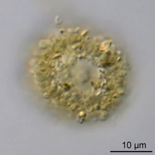
Empty shell, c. 33 µm
Drawing: from Penard (1902)
Filopodial network
Two cells from the same location
Frenzelina reniformis, just like Penards drawing above (l) and cell attached to the cover glass (r)
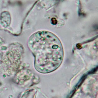
Dividing specimen, without test; kidney-shaped specimen without test
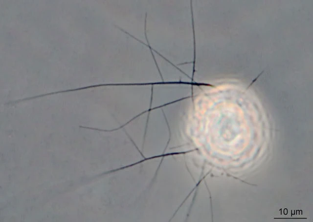
Small cylindrical aperture and bundle of filopodia
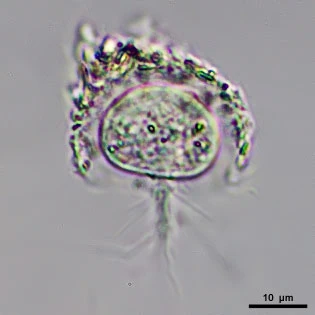
Pseudostomal view; lateral view
Cells about 24 µm, almost naked. Both cells from the same wet mount.
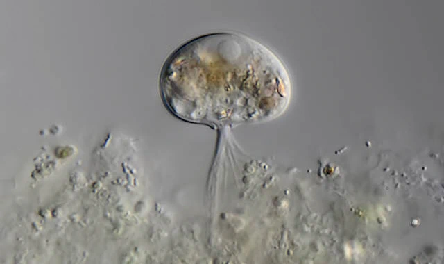
Frenzelina reniformis, with filopodia, cell c. 28 µm.
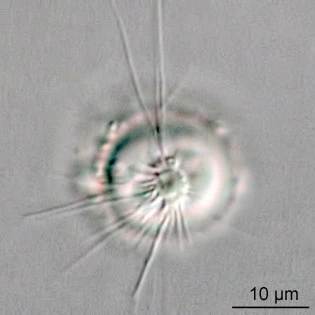
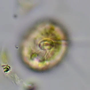
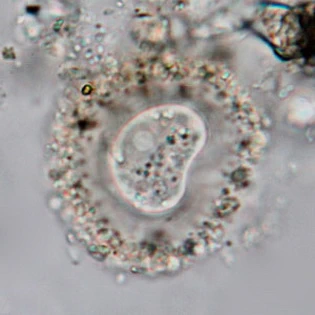
Shell almost a complete sphere, with small pseudostome
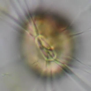
The same specimen, changing its aperture within one minute.
Frenzelina reniformis naked form
Frenzelina reniformis, nucleus visible
Naked specimen
Two naked specimens from the same wet-mount-culture. Notice the difference in size.
Frenzelina reniformis, 26 µm
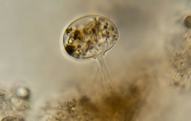
Frenzelina reniformis, with small tubular pseudostome; body 22 µm