Longitudinal division of a naked Frenzelina cell
December 16 I had several specimens of a large naked Frenzelina species in a wet mount which I had kept for over two weeks in de moisture chamber. I could observe a division of one specimen, at least the last part of it. It’s a longitudinal division, which means that the division starts at the “dorsal” side and ends with the division of the mouth or pseudostome.
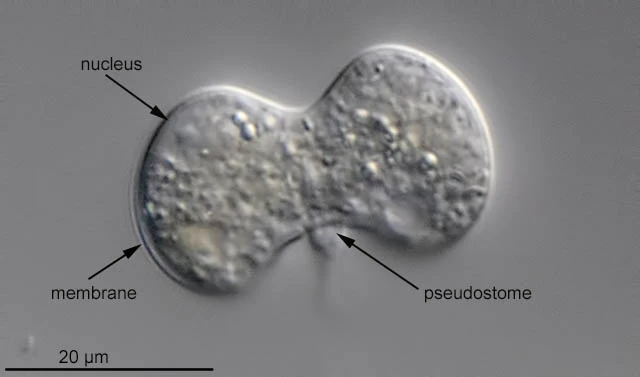
14:14:55
Beginning of observation; one of two nuclei is visible; there is one pseudostome.
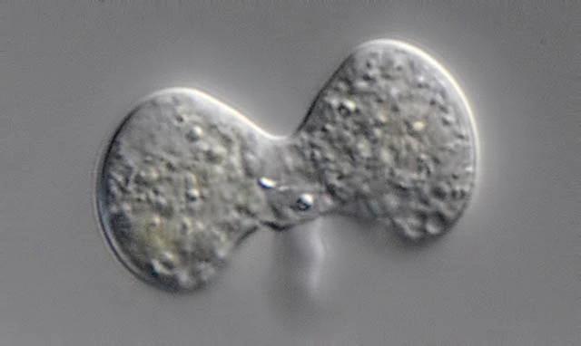
14:19:46
The “isthmus” between both cells gets smaller.

14:20:14
One thick lobopodium is present and one pseudostome.

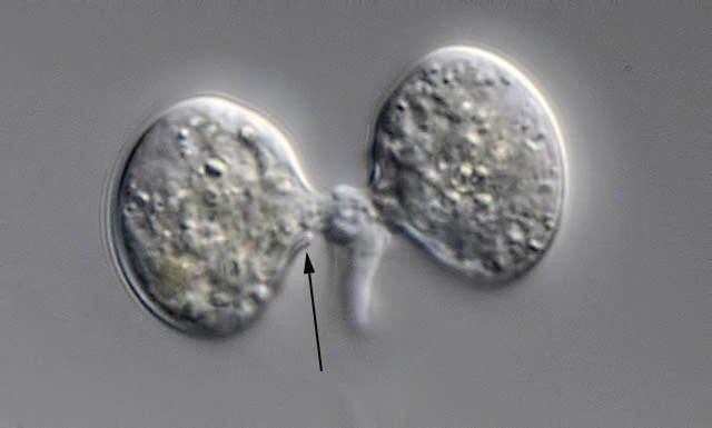
14:24:37
Each cell develops its own pseudostome. It’s not clear how the original pseudostome splits into two new ones.
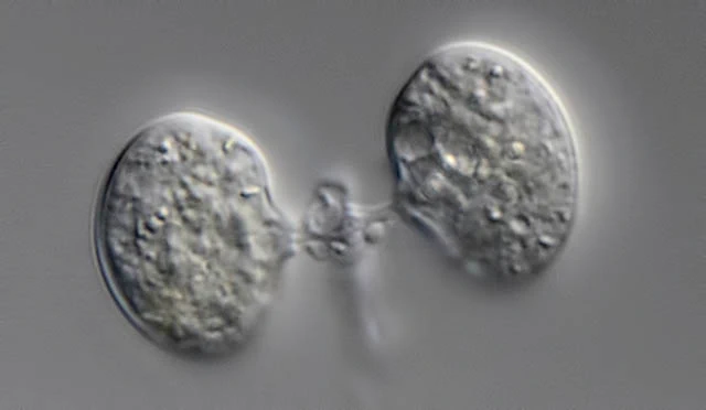
14:26:21
The arrow points to the collar of the new pseudostome.
14:30:05
Cells move farther away from each other.

14:32:53
Both cells are separated.