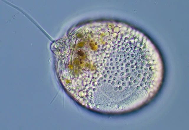
Lecythium granulatus, 82 µm
Lecythium granulatus (Schulze, 1875)
Diagnosis: test in lateral view broad elliptical or ovoid; in dorsal view circular; aperture flexible, with folds, often surrounded by a pliable collar; membane hyaline, colorless and flexible; cytoplasma usually with numerous fine granules; vesicular nucleus in the dorsal region; filopodia straight, branching and anastomising, sometimes extremely numerous; with veils between the base of some filopodia.
Dimensions: Literature 40-83 µm; my measurements 38-111 µm; nucleus c. 25 µm (hard to detect).
Ecology: I found this rather rare species in sediments of a ditch (Naardermeer) and the Spiegelplas, both in the Netherlands, and in the Weser river in Germany. This species is variable in size, and larger than other Lecythium-species which I have seen. Smaller species tend to have a nucleus with a more or less central nucleolar mass, while the nucleolar material in larger cells is scattered throughout the nucleus.
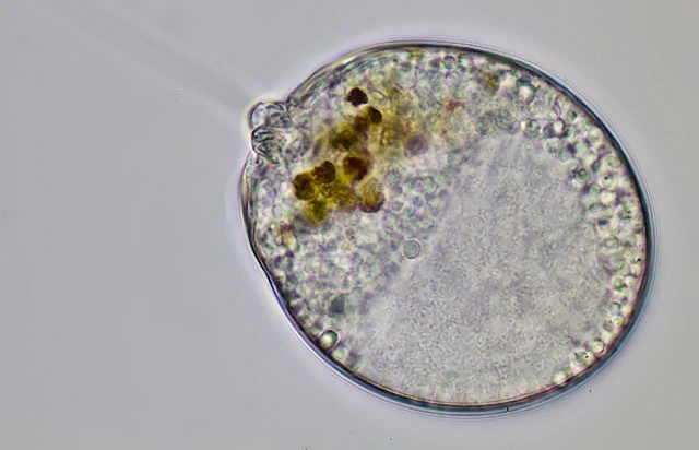
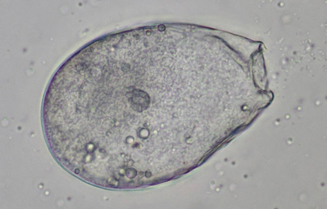
Cell with pseudostome (p) focused to the cover glass and surrounded by a pliable collar (c)the pseudostome is usually visible as a double ring
Two specimens from the same sample, 38 and 82 µm long.
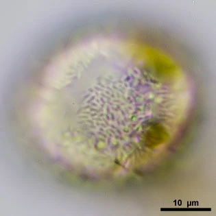
Specimen with typical granules; each granule bears a smaller granule.
The same specimen as above, with a large nucleolar part?
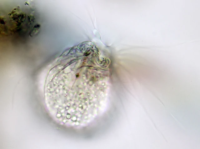
The same specimen as above, with large pliable and flexible aperture, surrounded by a kind of vacuolated collar.
Filopodia which branch and anastomose; between filopodia veils can be formed.
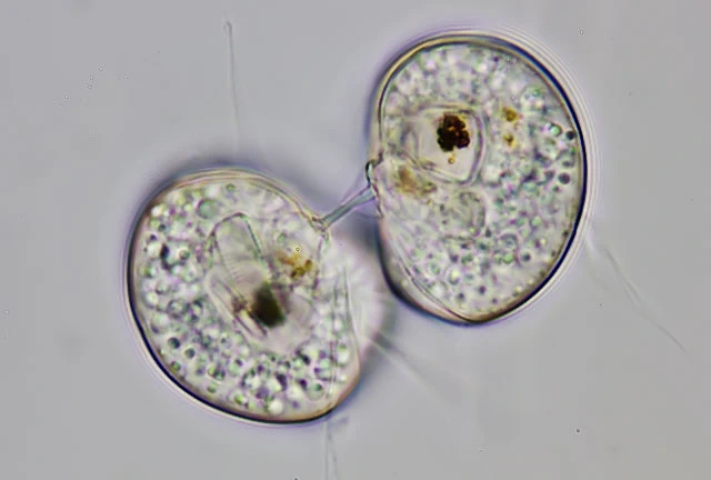
Two cell after division, just connected by a small string of plasm. Each cell c. 40 µm.
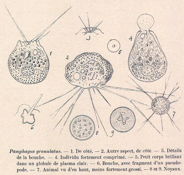
Lecythium granulatus, arrows indicate the nuclear membrane. The nucleus resembles the one drawn by Penard, see his Fig. 9 below, with a more or less central nucleolar mass. In this species the nucleus is very hard to detect. The above cell was compressed between slide and cover slip.
In DIC this surface structure became visible, bacteria? It looks if the bacteria adhere to the outer surface of the membrane (arrows).
Detail of filopodia and aperture
Lecythium granulatus
After Penard, in: Faune Rhizopodique du Bassin de Léman (1902).
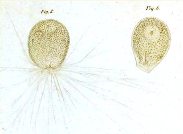
After Schulze, 1875
Empty membrane, 92 µm long; the living cell was 82 µm
Lecythium granulatus, 62 µm
Lecythium granulatus, 66 µm, with large funnel-shaped flexible collar.
Lecythium granulatus, 67 µm, with filopodia and large folds around the pseudostome.
Lecythium granulatus, with pseudostome and filopodia
Lecythium granulatus, 78 µm
Lecythium granulatus, Weser river, Germany – Phase contrast
Lecythium granulatus, Weser river, Germany – Phase contrast
Lecythium granulatus, Weser river, Germany – Phase contrast
Lecythium granulatus, Weser river, Germany – DIC