Microcorycia scutella Badewitz, 2004
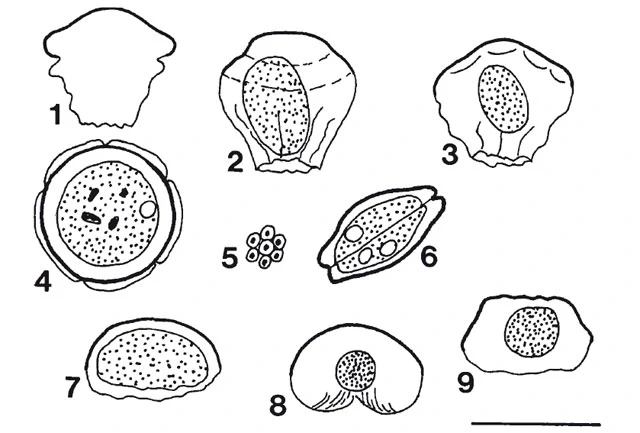
Diagnosis: Microcorycia with dome- or helmet-shaped upper test part which shows a delicate alveolar structure in the light microscope. Typical test shape of the genus Microcorycia. Upper test part dome- or helmet-shaped, sometimes with slight, irregular undulation; rounded in dorsal view; composed of rotund, irregularly arranged elements each with a dark centre (alveoli). Upper test part yellowish-brownish, lower test part hyaline. The cell body does not fill up the test. Cytoplasm is greyish and granulate. It contains one or two contractile vacuoles and food vacuoles with food particles (detritus?) and excreta. The cell is mononuclear. Pseudopodia and epipodia have not been observed.
Dimensions: mean = 50.8 (36-62) µm, n = 19.
Habitat: Xerophile mosses. Moss cushions from seven localities. Three epilithic mosses, four mosses from reed and tiled roofs. Germany.
Remarks: This species can be clearly distinguished from all Microcorycia species described so far. A delicate alveolar structure is also found with M. husvikensis (Beyens & Chardez 1997). However, M. scutella can be clearly distinguished from that species as it is bigger than M. husvikensis and its upper test part shows no divided margin.