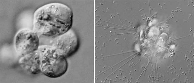
Genus Trachyrhizium Shiratori and Ishida, 2016
Diagnosis: Marine filose amoebae with thin smooth organic thecae. Filopodia thin, branching, and occasionally anastomosing, including small granules with bidirectional movement. Thecae consisting of two layers. Extrusomes present. Mitochondria with tubular cristae. Presence of Golgi apparatuses and microbodies.
Trachyrhizium urniformis Shiratori and Ishida, 2016
Diagnosis: Cells spherical, theca spherical with one wide circular aperture. Theca bilayered with an electron dense outer layer, and a slightly ambiguous and less dense inner layer. Extrusomes 0.4–0.5 µm in length and 0.15–0.2 µm in width, consisting of a spherical cap structure and electron dense cylinder that includes a less dense core.
Dimensions: Cells 7.5–17.6 µm in diameter.
Ecology: Marine sediments collected from northwest side of Agenashiku Island, Okinawa, Japan. Feeding on diatoms.