Acanthocystis pectinata Penard, 1889, emend. Siemensma & Roijackers, 1988
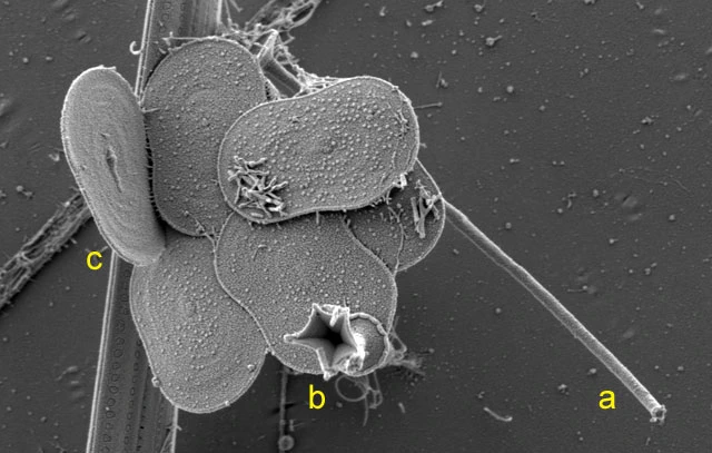
Diagnosis: Cell diameter 15-20 µm. Periplast with plate-scales and two kinds of spine-scales: short and long spine-scales. Short spine-scales all of the same length, 2.1-3.1 µm long, with a cylindrical, hollow and straight shaft, 0.25 µm thick; apex cup-shaped, 0.8-1.4 µm wide and with 4-6 long, diverging sharp teeth, interconnected by a thin membrane; basal plate 0.43-1.2 µm in diameter, conical, circular, with a fairly thick margin. Long spine-scales not abundantly present, 6.0-10.2 µm long with the apex terminating in 2-3 short teeth; no intermediate lengths between the short and the long spine-scales were observed. Plate-scales 1.6-2.5 X 1.3-1.6 µm, oblong to slightly ovoid with faintly concave sides with a broad marginal area ornamented with a pattern of small granules arranged in more or less concentric or radial rows. Similar plate-scales were observed by Dürrschmidt (1987a) and assigned to a new subspecies, A. pectinata ceylanica.
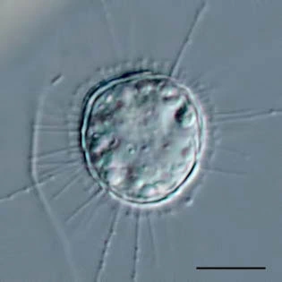
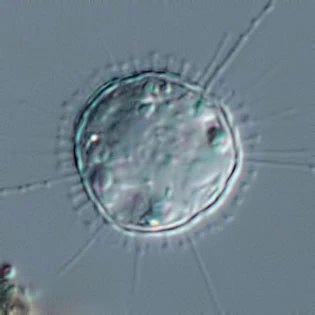
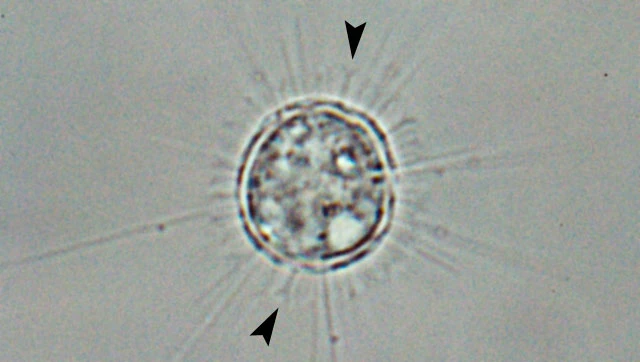
Remarks: I have found this species very often. Their habitus was in every aspect as described by Penard (1889, 1904) and Rainer (1968). The ring of short spine-scales is very helpful in identifying this species by light microscopy. In all specimens I detected a few thin, long spine-scales as reported by Rainer (1968). In vivo, these spine-scales are difficult to detect between the axopods. The diagnosis given by Penard (1889, 1904) and emended by Rainer (1968) is therefore sufficient to identify this species, even by light microscopy.