Darbyshirella spec.
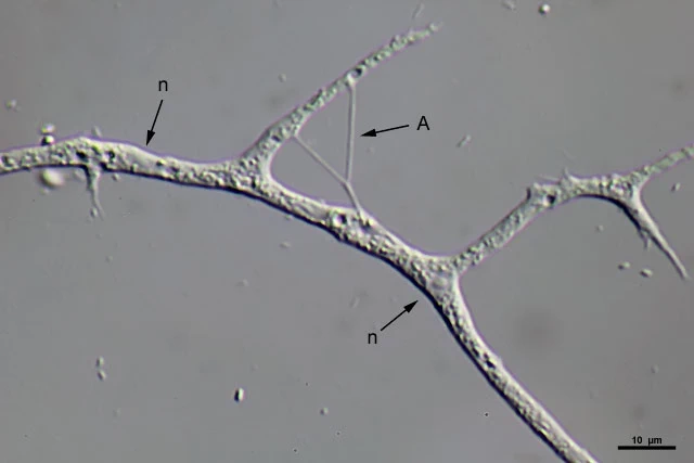
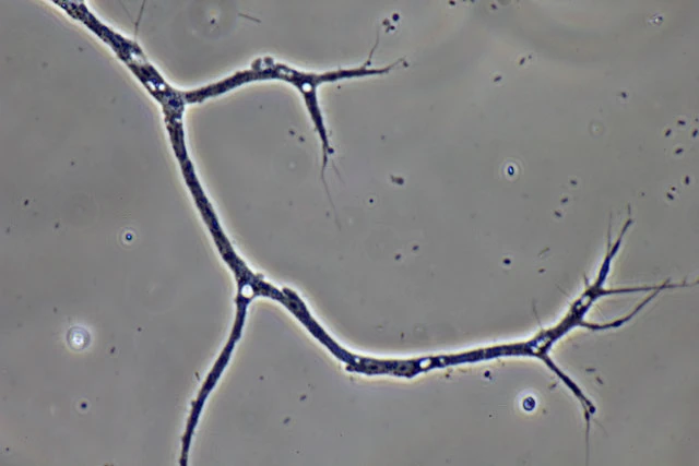
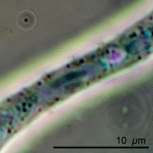
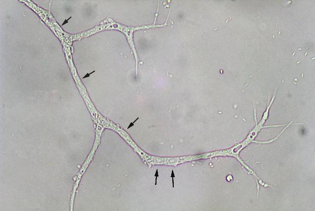
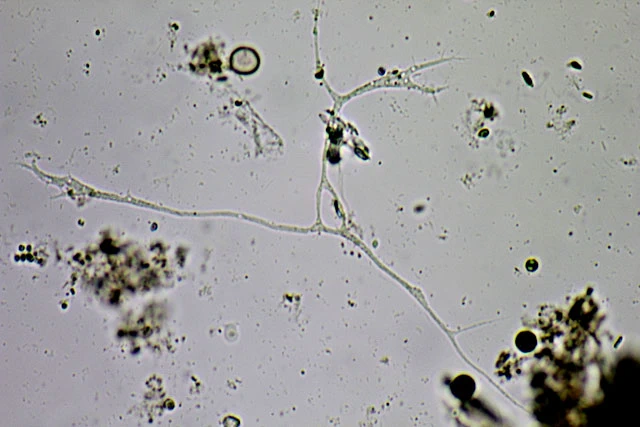
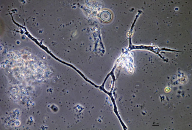
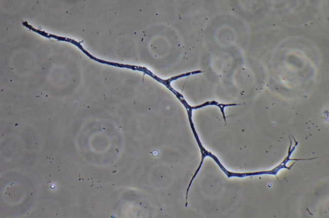
Diagnosis: Trophozoites ribbon-shaped, with continuously moving granular colorless cytoplasm, bidirectional; usually flattened and spreading on surfaces. Filopodia usually produced at the edges of the cell, often originating from a hyaline and very delicate fringe of cytoplasm. Filopodia long and thin, tapering, mostly unbranched, sometimes anastomosing. No membranosomes present on the pseudopodia. Cytoplasm with several contractile vacuoles; colorless granules, possibly corresponding to the membranosomes of Vampyrella. Several nuclei, about 10 pro specimen, hardly visible, vesicular, elongated elliptical or nearly spherical, with a central nucleolus, all shapes observed within one specimen. Cells usually stationary, moving very slowly over surfaces. No cysts observed.
Dimensions: my measurements: stretched body 300-350 µm; nuclei 4.1-6.8 µm.
Habitat: Fresh water, Sphagnum.
Remarks: The photomicrographs on this page are from two specimens which I found in a moist chamber preparation. They hardly didn’t move, only changed their filopodia. There was a continuing bidirectional streaming of granules. Each specimen had about ten nuclei, in shape varying from elliptical to elongated elliptical. The next day both specimens were gone, I couldn’t find any trace of them. The sample came from the Diepveen, a fen near Dwingeloo, The Netherlands.