Acramoeba dendroida Smirnov, Nassonova & Cavalier-Smith, 2008
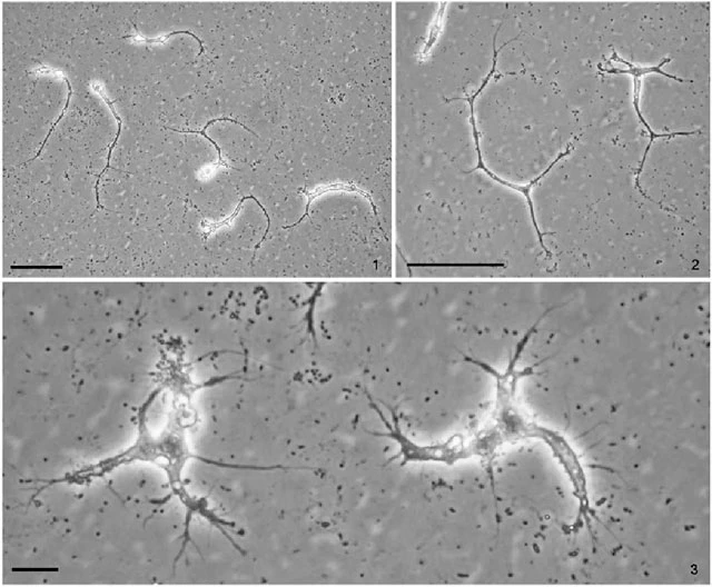
Diagnosis: Flattened, expanded and branched organism, up to 300 µm across; minimally mobile. Pseudopodia long, arm like; fine, ending up with fine, hyaline, non-granulated subpseudopodia. Uninucleate, vesicular nucleus 5-6 µm in diameter. Cysts rounded, single-walled, 25-35 µm in diameter. Simple life cycle consists of cyst and trophozoite stages.
Type strain: ATCC 50654. Type location: pond near Grand River, Grand Haven, MI. Freshwater.
Remarks: Smirnov et al. (2008) noted: “Trophozoites were of irregular shape, branched, with long arm-like pseudopodia. These pseudopodia branched, forming a tree-like pattern and subsequently decreased in width, ending with fine, hair-like hyaline subpseudopodia. Sometimes small islands of hyaloplasm were seen at the sites of branching. Separate subpseudopodia could be formed from any part of the cell as well. Trophozoites had very low mobility; virtually no visible movement of the main cell body was detectable; cytoplasmic flows in pseudopodia were hardly seen and only subpseudopodial activity was visible. These subpseudopodia very rarely fused, and in shape and structure resembled the pseudopodia of certain cercomonads, e.g. Cercomonas cometa. Some of them contained large refractive inclusions moving inside the cytoplasm, but the general pattern was very different from the typical granuloreticulopodial network of foraminifera and the movement of these inclusions was much slower. The size of the cell (across the longest dimension) varied from 50 to 350 µm, depending on its shape. The organism was uninucleate, with a single-vesicular nucleus 5-6 µm in diameter. Cysts were rounded, with a single, smooth wall, 25-35 µm in diameter. During 6 months of observation we saw only trophozoites and cysts in culture and nothing resembling plasmodia or flagella.”