Genus Amphizonella Greeff, 1866
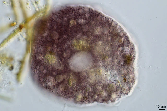
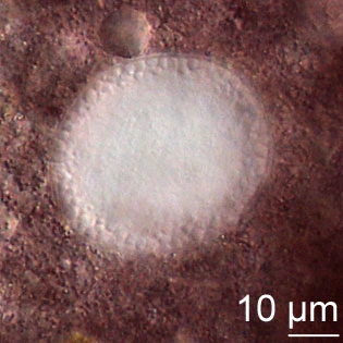
Diagnosis: body more or less spherical, surrounded by a flexible, bilayered pellicle; outer layer gelatinous, 8-12 µm thick with fine denticles, inner layer thin, chitinoid, undulates with internal movements, sac-like; pseudostome invaginated and variable; pseudopodia cylindrical, finely granular with rounded ends; movement slow; endoplasm clear, violet, yellow granules in purple vesicles; one or more ovular or spherical nuclei; the nuclear membrane is easily visible, the numerous nucleoli are mainly concentrated below the nuclear membrane. Contractile vacuoles 20-30 µm; no crystals; cysts reported.
One known species.
Amphizonella violacea Greeff, 1866
Diagnosis: as for the genus.
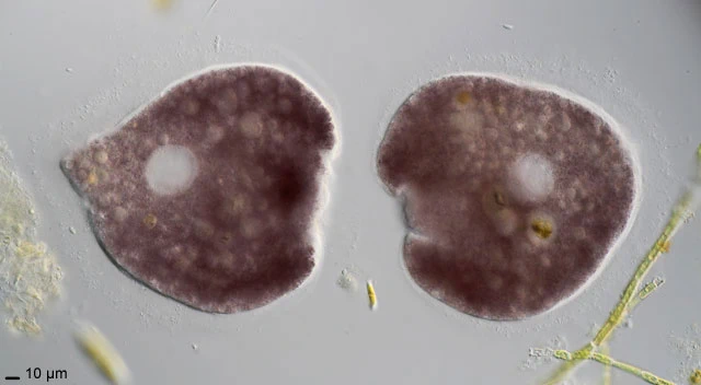
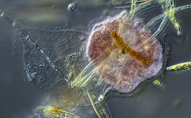
Non moving cells are usually roundish or oval, but can emit pseudopods. Moving amoebae adopt a more longish tongue or fan-like shape. Sometimes non moving amoebae take a stretched form with pseudopods at opposite ends. In contrast to previously published images the outer gelatinelike mucus layer is not always present. After excystation this layer is secreted de novo. Its thickness is variable and can reach up to 23 µm. Normally it contains numerous rod-shaped bacterial exobionts of unknown function. In SEM the outer surface is smooth. The untextured organic shell-wall is flexible and opens in a large not well defined aperture.
Dimensions: Literature 125-250 µm, nucleus up to 40 µm (Penard, 1906); Meisterfeld (2006): 75-558 µm. My measurements: 198-264 µm, nucleus 39.1-41.7 µm (n=8).
Habitat: Xerophilic mosses on roofs, also wet Sphagnum. Algivorous. I found this species in relatively large numbers between algae in a very shallow part of a freshwater pond (always present, at least from 2015-2025, Crailoo), also in Sphagnum in the Jura, France, 1986, and in dry mosses on a rock near Cervera de Pisuerga, in Northern Spain (2024).
Remarks: Meisterfeld and Badewitz (2006) collected this conspicuous, but often overlooked species, in xerophilic mosses from roofs in six localities in Germany. They found the overall shape much more variable than hitherto known and in one population of small specimen the violet color was lacking. The observed cells were uninucleate. Penard (1902) also observed one ovular nucleus per cell, but occasionally he found two or three nuclei in a cell.
References: Meisterfeld and Badewitz, 2006.