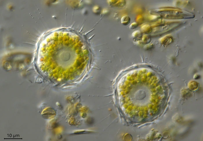
Choanocystis aculeata (Hertwig & Lesser, 1874) Siemensma & Roijackers, 1998
Basionym: Acanthocystis aculeata Hertwig & Lesser, 1874
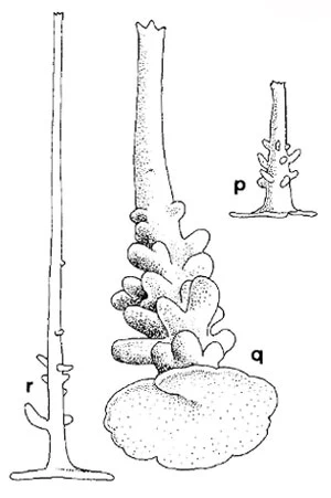
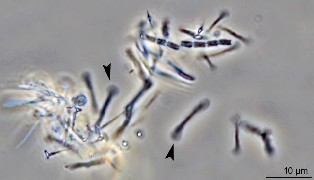
Diagnosis: Cell diameter 23-60 µm. Spine-scales 3.5-15 µm long, shaft cylindrical, hollow, straight or bent, tapering slightly to an open apex, ornamented with 5-11 diverging teeth; proximal part of the shaft commonly with a few or abundant nodules which are small or large digitate, sometimes even forked; shaft is set eccentrically, less commonly centrally, at the apex of a more or less deep incision of a cordate basal plate. Basal plate 2.0-3.2 µm in diameter and with irregular, often faintly lobed margins without any rim. In many cases, the edges of the incision overlap each other. Plate-scales 3.8-12.9 µm long and strongly variable in shape, small and elongated (rod-shaped) with more or less spatulate ends which are often widely expanded, resulting in a dumb-bell shape.
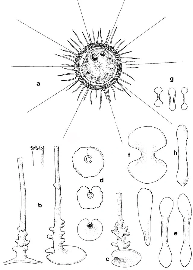
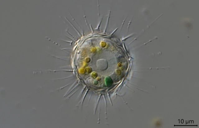
Fig. 1: a Living cell; b Spine-scales; c,d Basal plates; e-h Plate-scales or tangential scales – after Siemensma 1991
Remarks: I have seen spine-scales with large, even forked nodules and spine-scales with only faintly developed papillae. Both kinds of spine-scales are seldom present on the same specimen. There is no correlation, either qualitative or quantitative, between the shape of the plate-scales and the shape of the spine-scales. Individuals with dumb-bell shaped plate-scales seem to be more abundant than those with rod-like plate-scales.
Choanocystis aculeata is a well described species based on the shape of its plate-scales and is often recorded in literature (see Dürrschmidt 1985). The incised basal plate was mentioned previously by Siemensma (1981). Individuals with dumb-bell shaped plate-scales and papillate spine-scales were assigned by Nicholls (1983) to a new species, Acanthocystis serrata. Nicholls stated that Penard (1904) did not report the papillae and therefore Nicholls could not identify his specimen with Choanocystis aculeata. Hertwig and Lesser (1874) did not mention the papillae on the proximal part of the spine-scales, nor did Penard (1904). The first observations of the minute papillae were reported by Stern (1924), who speaks of “höckerige Unebenheiten”. Unaware of this short note, Siemensma (1981) gave drawings of these nodules. He recognized papillae after embedding spicules in Naphrax and using an oil-immersion objective. Penard, however, used Canada balsam, which has a lower refractive index, and he made his observations without an immersion objective (Deflandre 1958). If Penard did observe the papillae, it is not unlikely that he identified them as bacteria or considered them to be caused by distortion or aberration of the objective. The papillae are not always pronounced or even present (Croome 1986).
In the original light microscopic description this species deviates from most Acanthocystis species by its rod-shaped plate-scales (Hertwig and Lesser 1874; Penard 1904). A much larger variation was previously pointed out by Siemensma (1981) and confirmed by Dürrschmidt (1985) and Croome (1986). The form of the scales seems to depend upon environmental conditions, whereas there seems no differentiation in the shape of the spine-scales correlated to this reported variance.
One may conclude that Acanthocystis erinaceus Penard, 1889, falls within the range of variation of C. aculeata, though it is likely that Penard included the common A. erinaceoides in the description of A. erinaceus.


>