Equipment
Microscopes
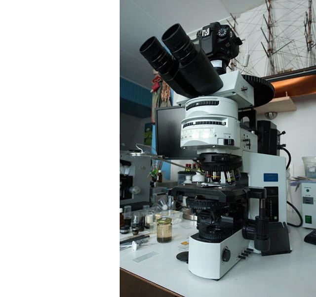
I examine my samples using an Olympus BX51 microscope equipped with Nomarski DIC, phase contrast, and fluorescence, along with various UPlanFl and UPlanApo objectives. I also utilize a Leitz Orthoplan microscope, which features plan apochromatic objectives, standard Köhler illumination, phase contrast, and a Smith T DIC system.
For observation, the material is transferred to standard microscope slides and covered with 24 x 32 mm coverslips. Specimen isolation is performed using custom-made, ultra-thin glass pipettes. These are created by heating a glass tube over a flame until pliable, then carefully pulling both ends to produce a fine pipette. This tool is used directly to collect amoebae, a process requiring considerable patience—and occasionally a fair amount of cursing!
Photomicrography and video
For scanning samples, cultures, and isolating specimens, I rely on a Nikon Diaphot inverted microscope equipped with phase contrast optics. For filming and photographing live specimens, I use a Caozhengwen MTR3 CMOS 26 MP camera on the BX51 microscope and a Canon 5D Mark II on the Diaphot. Image processing and measurements are performed with Adobe Photoshop and ToupView© software. Videos are captured using ToupView and later edited with Camtasia Studio for final presentation.
