Lacogromia cassipara Siemensma et al., 2017
morphotype A
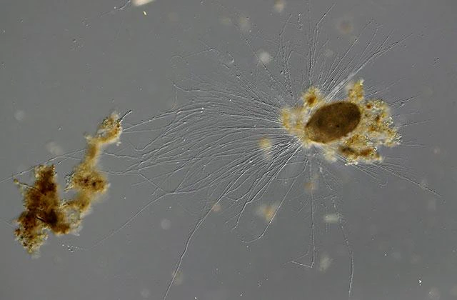
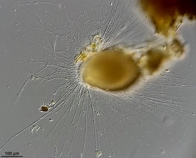
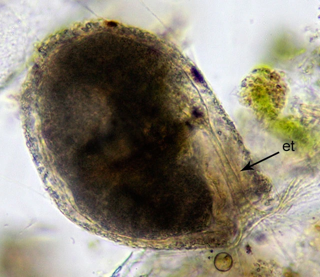
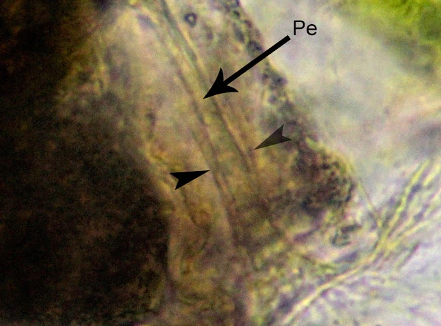
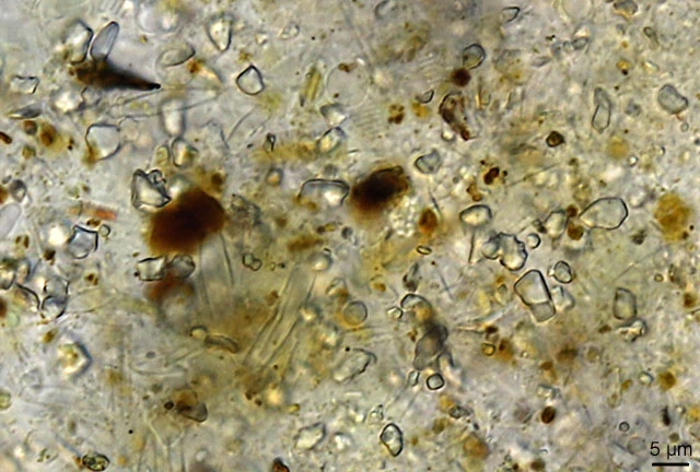
Diagnosis: Cells grey or brownish grey. Shell broadly ovoid or slightly pyriform, with usually one side stronger curved than the other side, giving the test an asymmetrical appearance. This stronger curved side, if present, not seldom bends slightly back towards the aperture. Aperture terminal, usually oblique. Cytoplasm separated from the aperture by the hyaline sheath, which surrounds the peduncle. The peduncle widens towards the aperture. Wall of the test composed of organic material embedded with very small xenosomes, mostly of siliceous origin, but also organic material can be present. Wall about 10 µm thick. Cells usually multinucleate, with up to 20 nuclei. Overall, cells with more nuclei have smaller nuclei. The nucleolar material is arranged in irregular rounded pieces distributed throughout the nucleus with slightly more nucleoli in the periphery. Granuloreticulopodia as characteristic for foraminifera.
Dimensions: Shell length 91-532 µm, mean 262 µm; test breadth 48-397 µm, mean 176 µm; L/B ratio 1.5; diameter of aperture 9-87 µm, mean 48 µm. Nuclei 16-56 µm.
Habitat: In organic sediment of stagnant oligotrophic and mesotrophic freshwater bodies.
This species was found in considerable numbers in March 2016 in a pond of the nature reserve Crailoo between the cities of Hilversum and Bussum in the central area of the Netherlands. This is a mesotrophic pond, about one meter deep with a sandy bottom and a thin layer of organic sediment. The precise sampling spot was located at 52.248385, 5.165926.
Remarks: Nuclei are difficult to observe when they are in the test. All those nuclei share the same structure: a large number of granular nucleoli, about 1.4 – 14.6 µm in diameter. Remarkable is that these structure changes completely when they are squeezed out of the test. Usually they are pressed through the smaller entosolonian tube and therefore damaged. Within a minute, the nucleoli disappear and a weakly granular nucleus remains.