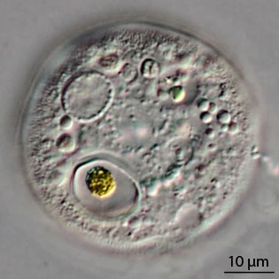
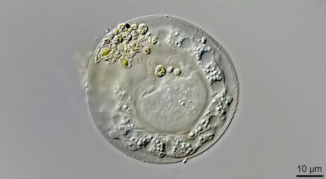
Microchlamys spec. n=nucleus Second specimen from Huizen
Microchlamys sp.
This species has a characteristic nucleus, broadly ovoid, where the nucleolar material is arranged peripherally. It was first discovered by Martin Kreutz, who published photomicrographs on a German forum in 2014. Shortly after I found several specimens in a small pond not far from the village of Huizen in the central area of the Netherlands. Food vacuoles contain small algae.
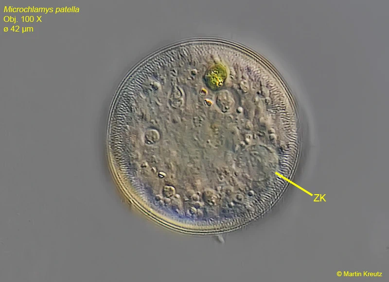
Dimensions: My measurements: shell Ø 47—50 µm, nucleus 10—10.9 µm; Martin Kreutz: shell Ø 42—64 µm.
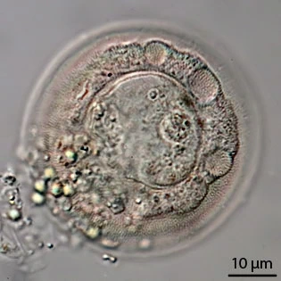
Microchlamys spec. with three contractile vacuoles; n=nucleus
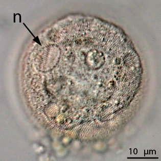
Microchlamys spec. Laegieskamp; notice the up to twelve contractile vacuoles in the periphery.
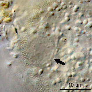
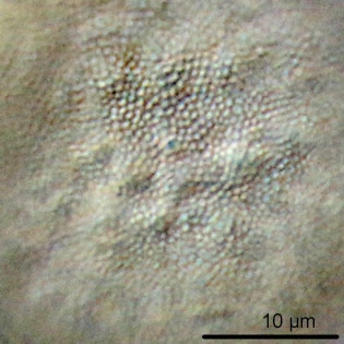
Microchlamys spec. nucleus arrowed – Laegieskamp, and at right structure of the dorsal side of the test.
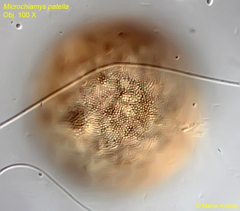
The photomicrographs below were made by Martin Kreutz, who sent them to me. He thought it was Microchlamys patella, but the arrangement of the nucleolar material is quite different from the central nucleolus as described for Microchlamys patella.
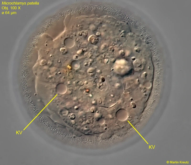
KV=contractile vacuole
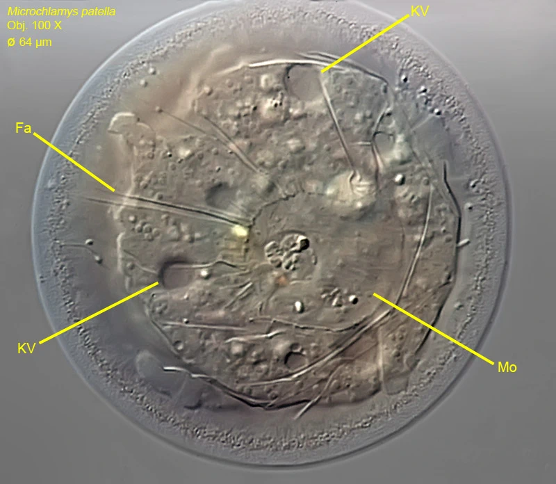
Fa=folds, KV= contractile vacuole, Mo=pseudostome
Mo=pseudostome, Fa=folds in the membrane
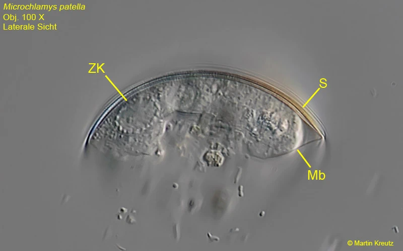
ZK=nucleus, S=shell, Mb=membrane
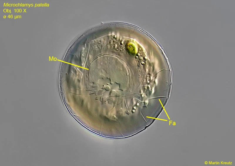
Mo=pseudostome, Fa=folds in the membrane