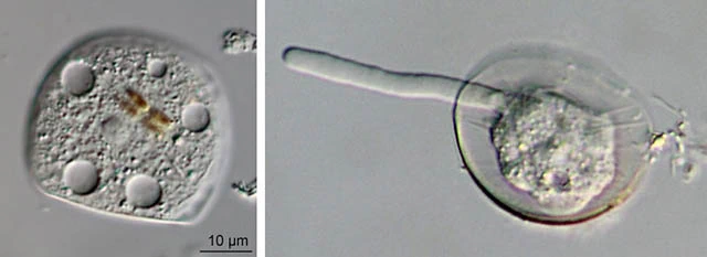
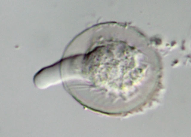
M. patella, with single lobopodium
Microchlamys patella (Claparède & Lachmann, 1859) Cockerell, 1911
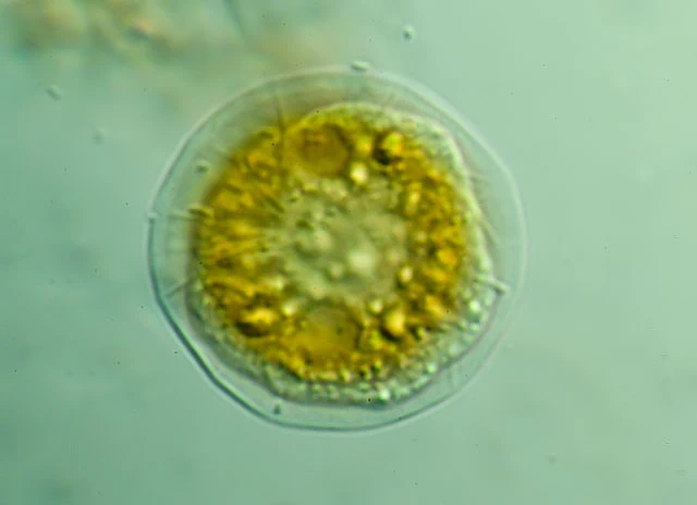
Diagnosis: Membrane or shell clear, yellow to brown, dish-shaped, flexible. Shell finely areolated, chitinoid, flexible and folded at the ventral side. Cell enclosed within a membranous sac which is fixed to the shell at intervals, but lost in empty shells. This membrane closes the shell from the ventral side and forms an aperture for the lobopodia. One vesicular nucleus, small crystals present.
Dimensions: 36—55 µm.
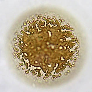
Remarks: It is not certain whether the photomicrographs on this page show Microchlamys patella or a Spumochlamys species. The photograph below shows a granulated pattern in the shell wall. This could be a Microchlamys.
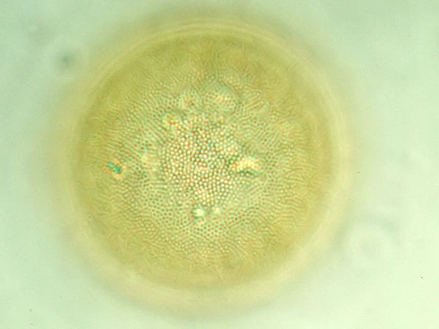
Microchlamys patella, surface structure.

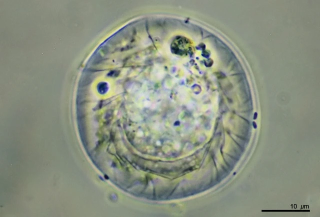
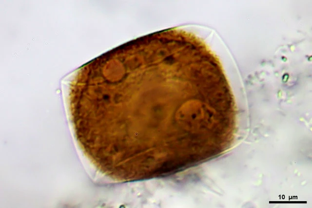
Pseudochlamys is a synonym for Microchlamys. Shell in cross section.
Notice the folds in the ventral membrane – Laegieskamp, The Netherlands

Nucleus (arrow) and nucleus with double nucleolus (right)

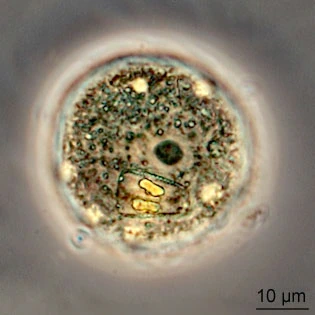
Both cells from a small pond in Gooilust, the Netherlands

Older membrane, 46 µm in diameter
Microchlamys patella, somewhat folded, in dry moss on a roof

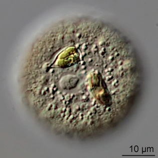
Microchlamys patella, 36 µm


Small specimen from a ciultured population, from Crailoo, The Netherlands


The same specimen as above