Velamentofex saxonensis Völcker and Clauß, 2020
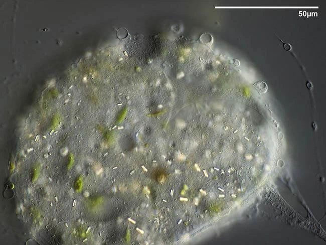
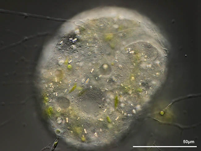
Diagnosis: Trophozoites ovoid, 30-95 μm, granuloreticulopodia up to 1000 μm long. with a hyaline, thin and extremely flexible membranous test sheathed with a broad layer of mucus-like material, that is covered with food particles and digestion residues. Nuclei ovular, 6.5-11.21 µm in diameter, with one central spherical nucleolus with a central lacuna and some irregularly shaped nucleoli located close to the nuclear membrane or with more and smaller granules scattered throughout the nucleus and one or two larger and more centrally arranged spherical nucleoli. Cytoplasm yellowish, with 5-12 contractile vacuoles in the periphery of the cell and with numerous refractive rod-shaped crystals, 1.0-4.2 µm long, or with numerous small refractive rod-shaped crystalline inclusions, attached to a spherical body, 1.4-3.6 µm in diameter. Cells multiplying inside the layer of mucus-like material, with daughter cells commonly staying inside this layer. Resting stages about 50 μm in diameter, formed inside the layer of mucus-like material.
Type locality. Submerged Sphagnum, peat bog area Rotes Wasser near Hormersdorf, Germany (50°38’58.25″N 12°54’16.44″E), Mai 2017.
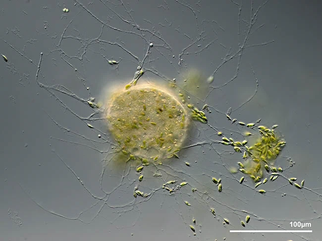
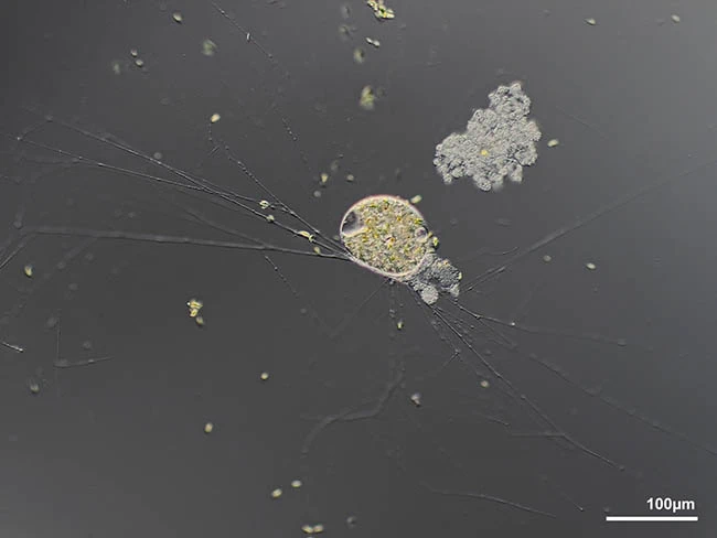
Description. All cells are ovoid with a hyaline membranous test which is very thin and extremely flexible, often with attached bacteria. The test is sheathed with a broad but not easily visible layer of mucus-like material, that is covered with food particles and digestion residues, which makes it often difficult to recognize the cell. Cytoplasm yellowish, with numerous food vacuoles. Some specimens contain numerous refractive rod-shaped crystals that are somewhat narrowed in the middle part, while other specimens contain numerous small refractive rod-shaped crystalline inclusions, each attached to a spherical body. Some specimens contain nuclei with one central spherical nucleolus with a central lacuna and some irregularly shaped nucleoli located close to the nuclear membrane. These nuclei are 9.3-11.21 µm in diameter. Other specimens contain smaller nuclei, 6.5-8.1 µm in diameter, with more and smaller granules scattered throughout the nucleus and one or two larger and more centrally arranged spherical nucleoli. The pseudopodial network may cover an area up to 2000 μm or even larger in diameter. Cells move slowly along a polar extended relatively thick pseudopodium. Stationary cells build an envelope of algae and digestion residues.
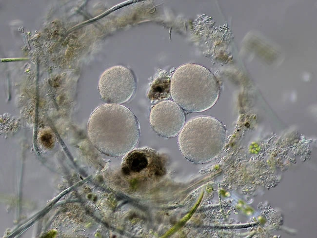
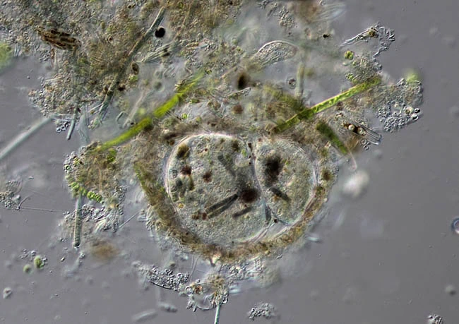
Cells multiply inside the layer of mucus-like material, with daughter cells commonly staying inside this layer. Daughter cells can also move out of the layer along the peduncle and migrate as small long bulges along the pseudopodial network. Under culture conditions actively feeding cells sometimes become extremely large and shapeless.
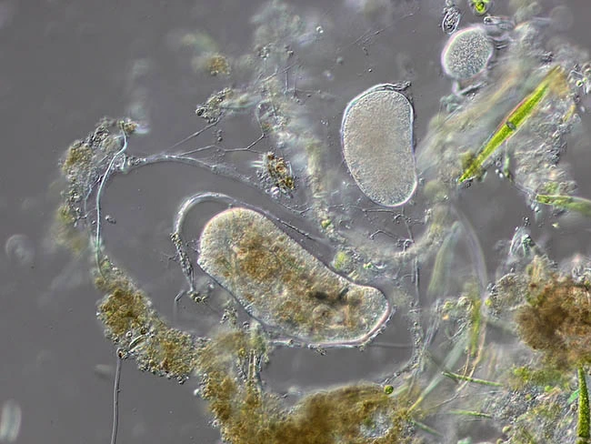
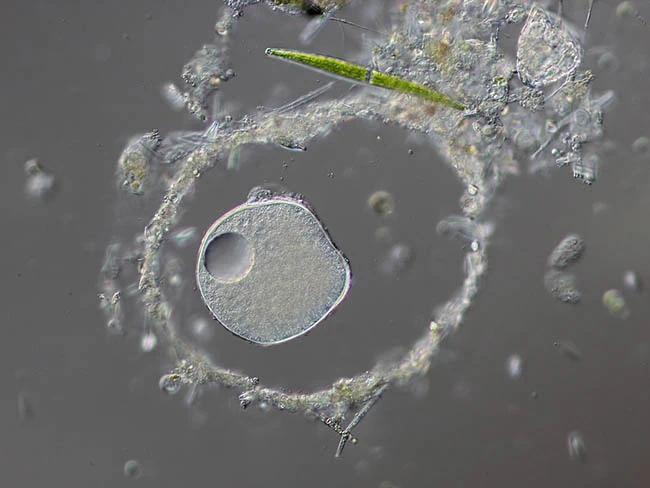
Resting stages are formed inside the layer of mucus-like material, first with a thin layer but after some days with a distinctly thicker and multilayered envelope of mucus-like material. Feeds on coccale algae, cyanobacteria, diatoms, rotifers and yeast.
Remarks: This species differs from V. berolinensis in the structure of the nuclei (a central nucleolus and some smaller peripheral nucleoli vs. a completely fine-grained nucleus) and their size (6.5-11.2 µm vs. about 11.5 µm). It differs from V. tyrolensis in the structure of the nuclei (a central nucleolus and some smaller peripheral nucleoli vs. some small nucleoli scattered through the nucleus).