Phryganella acropodia (Hertwig & Lesser, 1874)
Basionym: Difflugia acropodia Hertwig & Lesser, 1874
Synonym: Phryganella hemisphaerica Penard, 1890
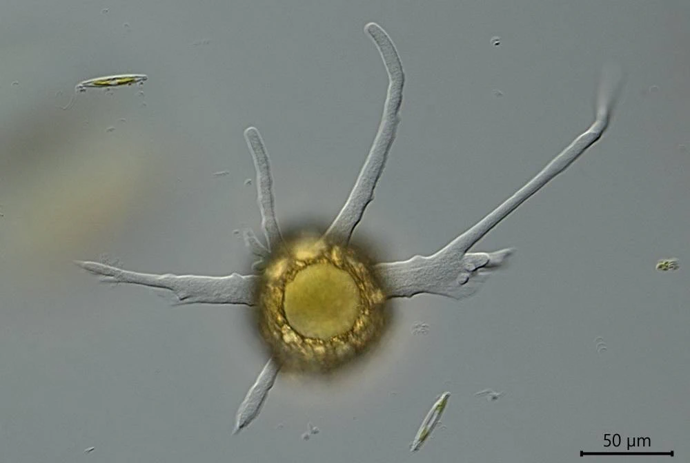
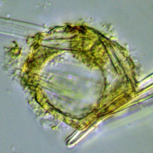
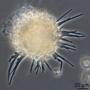
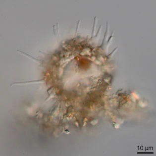
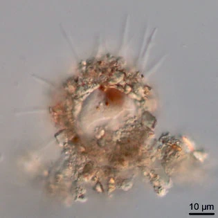
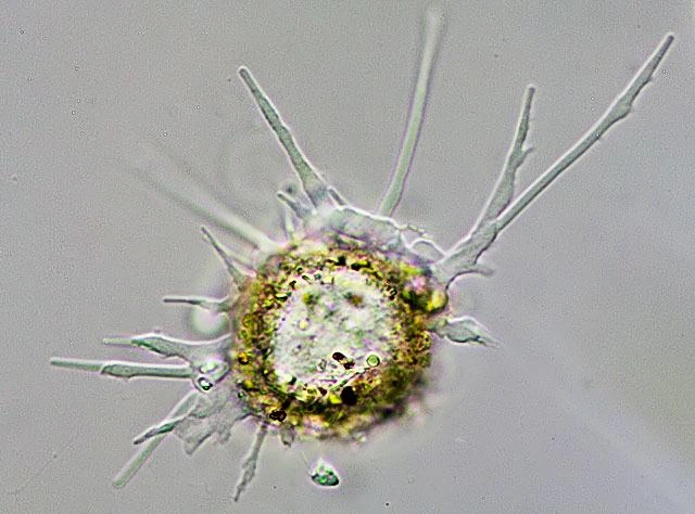
Diagnosis: Shell circular in apertural view, approximately hemispherical in shape and semi-circular in lateral view, with a small slightly invaginated rim; yellowish or brownish, semi-transparent, and covered with sand-grains and scales; in front view sharply pointed pseudopodia radiating; colorless endoplasm usually with chlorophyllous bodies; shell composed mainly of small or medium sized mineral grains which are incorporated into the matrix to give a regular, smooth, outline around the apertural and central regions but at the aboral extremity more angular grains may be incorporated; organic cement is only occasionally visible as small strands between adjacent particles; aperture large, circular.
Dimensions: 30-50 (in literature); 48-57 µm in diameter (Dutch sequenced strain, in Dumack et al., 2018).
Ecology: Freshwater, different water types; common.
Remarks: Examination of portions from broken specimens reveal that organic cement lines the inner surface which is always smooth and featureless. However, the outer face of the wall is composed of a small open network into or onto which the mineral particles of the surface are either bound or embedded. At the aperture this inner network is curved over so that it becomes, for the extend of the organic border, the outer region with mineral particles sandwiched between layers of organic network. It is noticeable that in these agglutinate shells the apertural region is thickened by incorporation of mineral particles between building units, but at the fundus only a thin layer of units lines the inner surface onto which a thick layer of grains is attached.
Penard (1902) erected genus Phryganella to accommodate those hemispherical specimens of Difflugia which had pseudopodia that varied from broad lobate to narrow digitate with pointed extremities, and included D. acropodia as a synonym of his P. hemisphaerica (Penard, 1890). Since then, Cash & Hopkinson (1909) have corrected the designation of the species and it has become P. acropodia. As these latter authors pointed out, one of the difficulties in identifying empty shells of P. acropodia is its similarity to empty shells of Difflugia globulus and Pseudodifflugia gracilis which all share roughly similar dimensions and shell architecture. The composition of the shell matrix in these species also varies, with soil specimens being predominantly composed of mineral grains, whereas those in Sphagnum and freshwater are composed mainly of siliceous shell plates and whole or broken portions of diatom frustules.
References: Dumack, K., Görzena, D., González-Miguéns, R., Siemensma, F., Lahr, D.J.G., Lara, E., Bonkowski, M. 2018. Molecular investigation of Phryganella acropodia Hertwig et Lesser, 1874 (Arcellinida, Amoebozoa). Eur. J. Protozool., https://doi.org/10.1016/j.ejop.2020.125707.


Original description in Hertwig and Lesser, 1874
Difflugia acropodia nov. spec.
Translated from German into English:
“The shell of this not very common organism is rounded and has an average diameter of 0.05 mm. It consists of a homogeneous, translucent membrane through which—if it is not too densely covered by foreign particles—one can see the shell opening of the animal as it crawls across the slide. The foreign particles consist mostly of bits of silica or small diatom frustules and lie on the membrane of the shell like fieldstones on shingle roofs: usually, like those, at some distance from one another, but often also so densely packed that they completely cover the actual shell.
The pseudopodia of our species differ from the blunt, finger‑shaped extensions of most other *Difflugia* by their pointed ends, which recur in all their manifold forms. Broad plates of homogeneous protoplasm end, at some distance from the pseudopodial opening, in irregularly shaped outgrowths and lobes with extremely characteristic, sharply jagged contours. In addition, wavy‑edged, generally lanceolate extensions arise directly from the body, very similar to the pseudopodia of an Actinosphaerium, except that—unlike those—they do not contain granules within. These actinophrys‑like homogeneous extensions may branch repeatedly, like those of the Monothalamia Rhizopoda; they also have a strong tendency for their tips to fuse with one another. As they flatten and spread out into surface‑like expansions, they gradually transform into the broad, jagged‑contoured plates described above. This occurs because the protoplasm rises from the base of the branched pseudopodia like a kind of webbing, or because pseudopodia fuse with one another and the resulting slit‑shaped gaps disappear as the sarcode rapidly advances from all sides.
Throughout all these changes, the movement is extraordinarily lively. When the Difflugia lets its sharply indented, pointed pseudopodia flow outward, it gives the impression as though a liquid were being poured from the shell and spreading rapidly across the slide. With the same liveliness, the contours change constantly: extensions are withdrawn and new ones formed.
Sometimes one also sees blunt, club‑shaped extensions among the pointed ones described. These are always present in small numbers and exist only briefly. Like the broad, finger‑shaped pseudopodia that occasionally appear among the pointed and fine ones in Vampyrella, we interpret them here as signs of discomfort—caused when, through evaporation of the water, the coverslip presses too heavily on the specimen being observed, or when the gas content of the water has changed.
From this description it is clear that the pseudopodia of D. acropodia possess a peculiar mixture of characteristics that otherwise do not occur at all, or only exceptionally, within the group of the Lobosa. Alongside thin and broad protoplasmic plates, there are pointed, almost thread‑like, branched and anastomosing extensions—and all this in a species belonging to a genus otherwise distinguished by its typical simple, finger‑shaped or lobed pseudopodia. Without a sharp boundary, these forms lead from the blunt pseudopodia of the Lobosa to the pointed ones of the Rhizopoda. Here we have further evidence for our view that, given the diversity of forms taken by the protoplasm functioning as an organ of locomotion—from the amoeboid flowing of the body and the lobed extensions of the Arcellinae to the richly branched pseudopodia of the Foraminifera, connected into fine networks—the intermediate stages are not lacking.
Despite the seemingly favorable shell conditions, we were unable to gain insight into the internal structure of the body, because the relative smallness of the animal made the otherwise customary and most suitable method for studying the organization of Difflugia—that of crushing—difficult.”
—
Below the description of Phryganella haemispharica by Penard, 1890:

Phryganella hemisphaerica Penard, 1890
Diagnosis: Penard (1902):
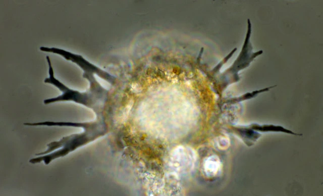
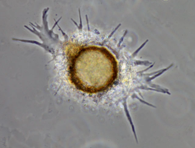
“Shell hemispherical, yellowish or brownish, rather transparent, formed of a mixture—highly variable in proportion—of amorphous siliceous scales, quartz grains, diatoms, and often composed almost entirely of the frustules of these latter organisms. On the lower surface, the shell wall bends sharply and opens into a generally very large aperture, reaching half or more of the total diameter (fig. 4, above), and only rarely smaller (fig. 1 and 3 above, where the aperture diameter has been drawn too small). This aperture is not invaginated and shows no trace of a collar; it is bordered by the covering scales, which are sometimes larger here than elsewhere and arranged in a certain order (fig. 4). The nucleus is single, round, generally eccentric, and contains a large granular nucleolus (fig. 5).
The pseudopodia show a strong resemblance to those of Phryganella nidulus, while at the same time approaching more closely those of the genus Pseudodifflugia: sometimes linear and thin (fig. 3), sometimes a little broader, sometimes straight and sometimes branched (fig. 1), or spread at their base into a wide sheet which itself divides into digitiform extensions (fig. 4). In general, one may say that when the animal is moving rapidly and has not been disturbed for some time, the pseudopods are all linear and very narrow; one may also add that in this species the movements are extremely lively and the deformation of the arms rapid.”
Dimensions: Penard (1902): Shell diameter 41-55 µm, with smaller specimens 25–40 µm occurring in other locations.