Vampyrella pendula Cienkowski, 1865
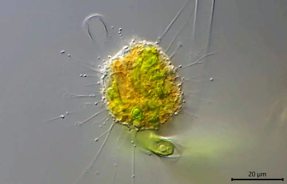
Diagnosis: Cells compact, often spherical, rarely deviating in shape; size of young trophozoites about 20–30 um. Cell body with radiating pseudopodia, occasionally accumulating at the frontal edge during movement. Intense orange coloration of the central cell body, periphery and pseudopodia colorless. Cells float freely in the water, or perform stalking, paddling movements. Pseudopodia very long (up to 50 um) and thin, tapering, branched or unbranched; hyaloplasmatic; more or less equally distributed over the cell, sometimes in tufts. No membranosomes present on the pseudopodia, contrasting V. lateritia. Rarely formation of broad, claviform pseudopodia occurs, possibly related to pathological conditions. Plasmodia. Cell fusion not yet observed.
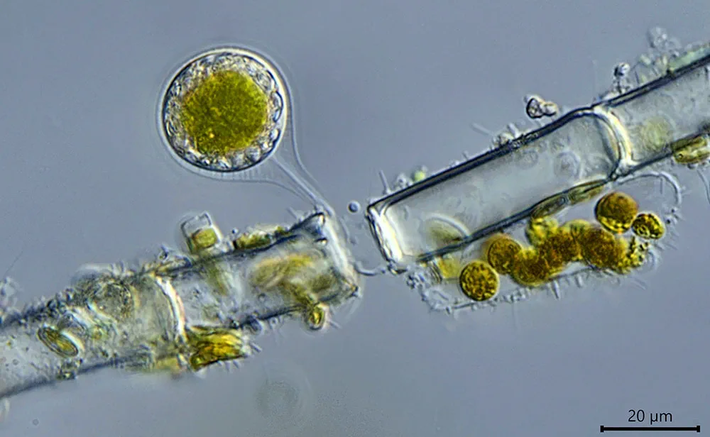
Digestive cyst outline spherical, or very slightly elliptical; size about 25 mm in diameter; attached to food alga with outer cyst envelope drawn out into a very characteristic hollow stalk. Three cyst envelopes: outer envelope delicate, with even surface; shape pyriform; distally attached within an empty algal cell and thus functioning as stalk. The next inner envelope delicate, sometimes hardly visible; seems to be connected to the outer by very faint strand-like structures. The innermost cyst envelope is comparatively prominent, with even surface; covering the cell body tightly before hatching; a hyaline strand seems to be connected to the innermost cyst envelope and runs within the two outer cyst envelopes to the distal attachment site of the cyst stalk. Young cyst stages with voluminous green content covered by colorless peripheral cytoplasm, exhibiting a very faint, transparent sheath (maybe mucilage) rarely visible in DIC; during digestion the cytoplasm turns orange-red; mature cysts contain a central, brown food remnant, which is left behind in the empty cyst after hatching. Resting cysts observed.
Feeding behavior: In general resembling V. lateritia; until now V. pendula not observed to feed successively on several algal cells. Proven food organisms: Oedogonium stellatum, Oedogonium cardiacum; V. pendula in the natural sample was feeding on Oedogonium sp.
(Diagnosis after Hess S, Sausen N, Melkonian M (2012) Shedding Light on Vampires: The Phylogeny of Vampyrellid Amoebae Revisited. PLoS ONE 7(2): e31165. doi:10.1371/journal.pone.0031165)