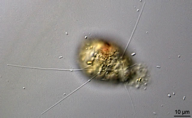
Capsellina sp.?
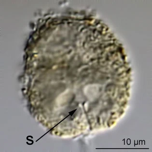
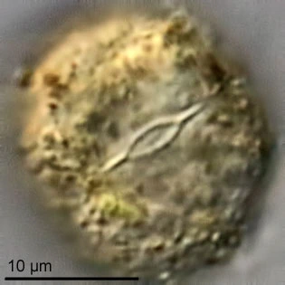
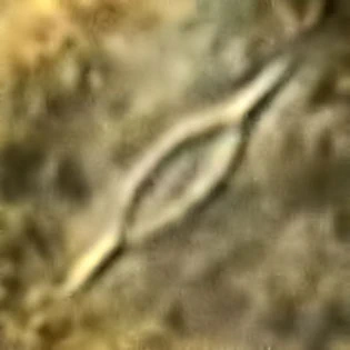
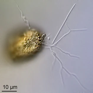
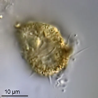
I found this species in shallow fresh water, Crailoo (2015) and it multiplied well during some days in moist chamber preparations. Its pseudostome is remarkable. The surface shows a slit-like invagination which goes down into the cell for about 5 µm, where it ends in an opening with a thickened rim. This opening can be closed. Usually it is open and has a shape something like =O= (see photomicrographs below). This pseudostomal structure, as I call it, is very distinct, but not always clearly visible, as it can be obscured by other material. The cell is surrounded by a membrane which is covered with minute xenosomes. The plasmabody fills the membrane completely. There is one vesicular nucleus with a relatively small central nucleolus which shows one or more lacuna.
Dimensions: cell 36-48 µm; nucleus 9-11 µm; inner pseudostomal structure c. 8-10 µm.