Diplophrys archeri Barker, 1868
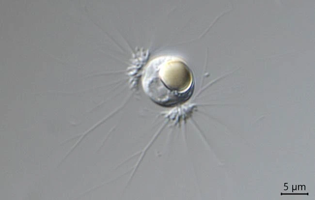
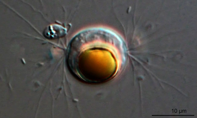
Diagnosis: Cell spherical or somewhat elliptical, covered with a thin, hyaline and colorless membrane; two circular pseudostomes situated at two more or less opposite poles; cytoplasm colorless, transparent, finely granulated, filling the envelope except near the two pseudostomes; a single nucleus with a nucleolus; one or more contractile vacuoles; one large, or two or three small, grayish, yellow, red or orange colored, oil-like globules present; pseudopodia extremely attenuate, in locomotive cells radiating, straight or dichotomously branched, in stationary cells curved and branched and attached to the substrate; reproduction by fission or tetrad division. Young cells frequently aggregated into colonies which form circular masses about 30-60 µm in diameter or more; from the periphery of which slender pseudopodia radiate.
Dimensions: solitary cells 8-20 µm in diameter, young cells in a colony 2-4 µm in diameter.
Habitat: solitary or in groups; on submerged plants or in debris in fresh water. Common in different types of water.