Mastigamoeba aspera Schulze, 1875
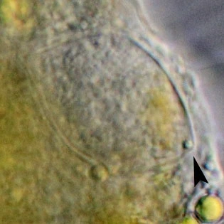
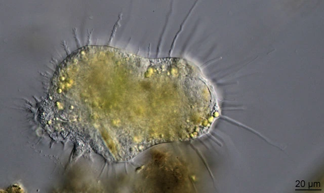
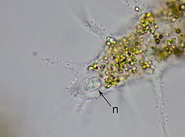
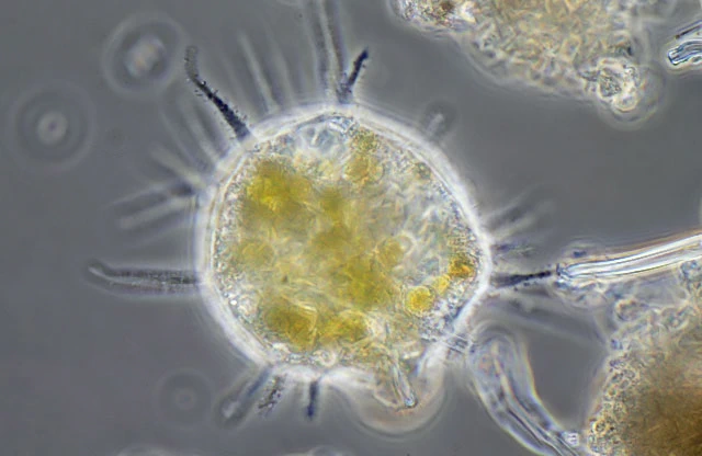
Diagnosis: Body semispherical or oval; during locomotion oval or pyriform; one motile cilium located at the anterior end of mononuclear forms; usually with a bulbous uroid; numerous conical or finger-shaped hyaline pseudopodia; outer surface usually inhabited by numerous rod-shaped ectobiotic bacteria; vesicular nucleus with a large central spherical nucleolus; cilium closely connected morphologically with the nucleus.
Dimensions: 50-250 long by about 50-100 µm broad, nucleus 16-20 µm, cilia c. 100 µm.
Ecology: in the ooze of ponds and ditches. It has also been found in brackish water (Carlos Gomez Revello, Uruguay, pers. comm., 2016)
Remarks: Both mononuclear cells with cilia and multinuclear cilium-free individuals have been observed. The base of the cilium is represented by a single kinetosome, from which radial microtubules and a lateral rootlet pass out into the cytoplasm. At the base of the kinetosome, there is a compact center of organization of microtubules (COMT), in which there are immersed bases of the nuclear cone microtubules participating in formation of karyomastigont. The structure of the cilium axoneme corresponds to the formula 9(2)+2. The main volume of the cytoplasm is occupied with digestive vacuoles. In addition, the cells contain numerous light-reflecting granules, as well as glycogen granules. Mitochondria, dictyosomes of the Golgi apparatus, and microbodies are not revealed (Chystyakova et al, 2012).