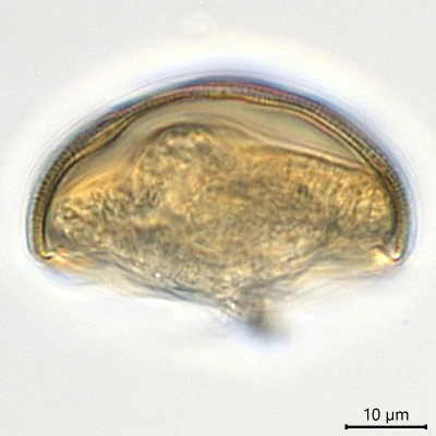
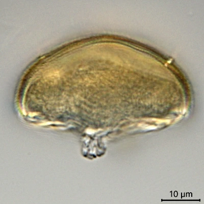
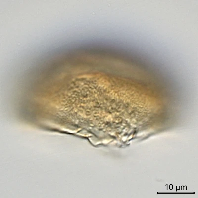
Microcorycia spec., the same cell in lateral view
Microcorycia spec.
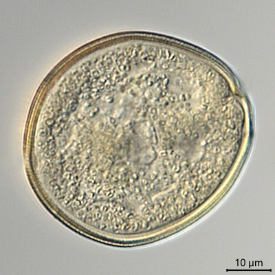
Diagnosis: Shell relatively high compared with all described species. The diameter is twice the height. The shell is finely granulated over its whole dorsal part in contrary to the other species which have a smoother non-granulated or much more finely granulated zone along the border between ventral and dorsal region. The ventral part was closed by a wrinkled membrane. The only cell that was observed, seemed to be dead, without any visible nucleus.
Dimensions: Shell Ø 48 µm, c. 22 µm high (n=1).
Ecology: Moss on fallen tree in marsh, Naardermeer, Netherlands (2019).
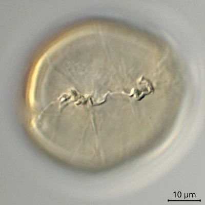
Microcorycia spec., oral view
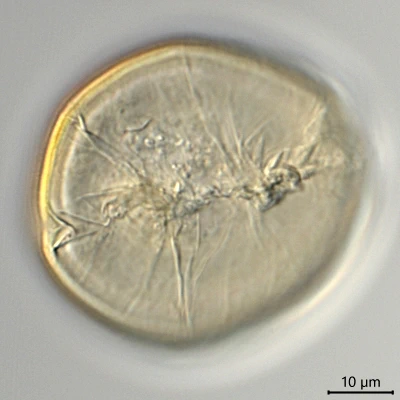
Microcorycia spec., dorsal and lateral view