Amoeba leningradensis Page & Kalinina, 1984
Diagnosis:
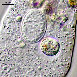
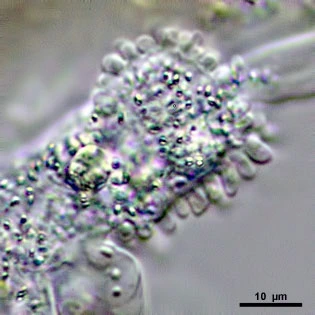
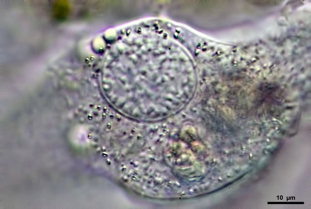
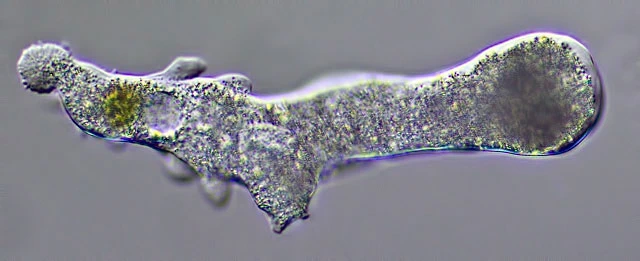
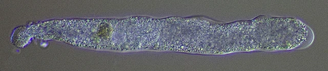
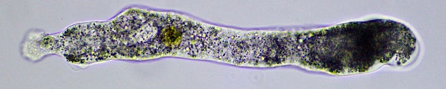
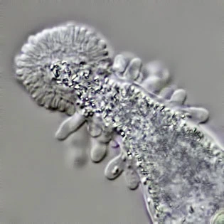
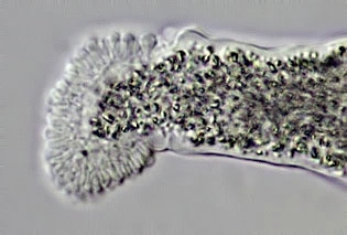
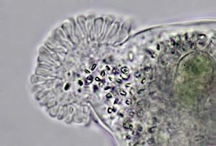
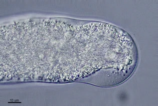
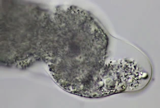
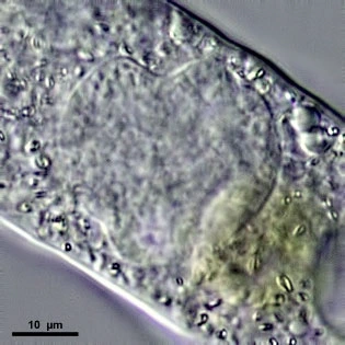
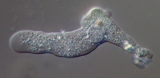
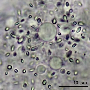
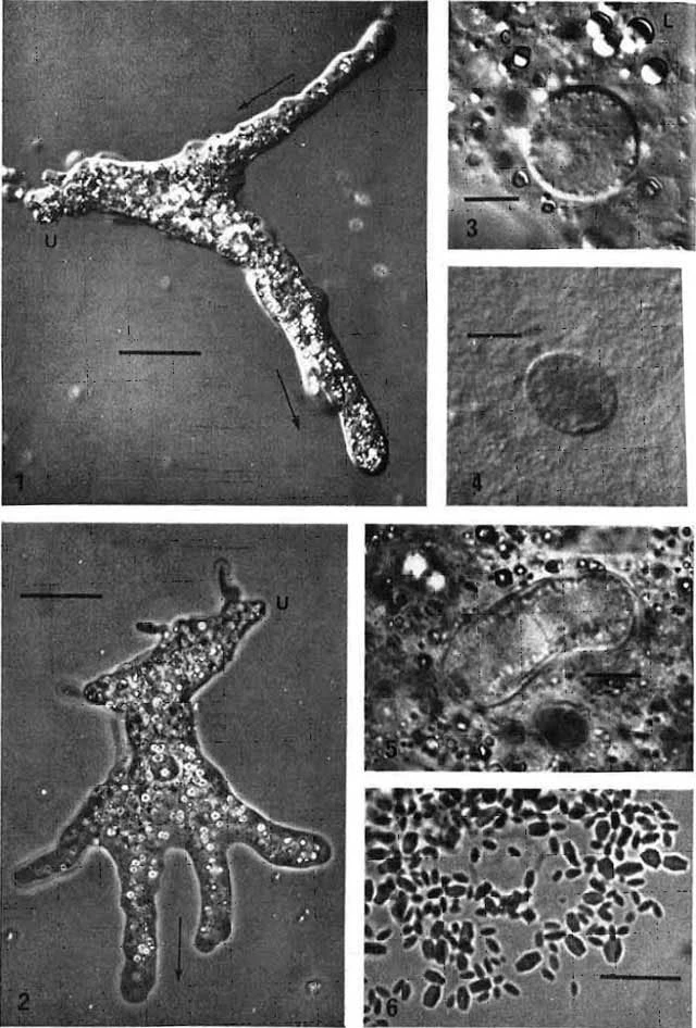
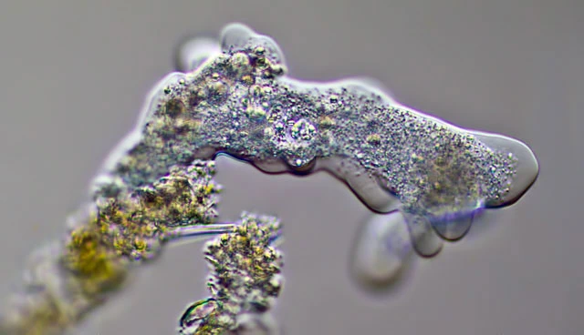
Locomotive form often polypodial, 160–550 µm long (mean ~360 µm). Hyaline cap less prominent than in A. proteus. Nucleus spherical but often slightly compressed, frequently appearing ovoid; greatest diameter 21–29 µm (mean 25 µm). Cytoplasm with numerous crystals, mostly truncate bipyramids. Uroid coarsely morulate or knobby, occasionally containing trailing remnants of pseudopodia that have passed to the posterior end.
Plasma membrane covered by an amorphous coat 13–20 nm thick. Nuclear envelope with an internal fibrous lamina differentiated into hexagonal prisms; lamina usually 400–550 nm thick, inner diameter of each prism ~165 nm. Nucleolar material concentrated around the nuclear periphery but also present internally. Golgi bodies composed of about four flattened saccules with associated vesicles, sometimes possibly of vesicles alone. Nucleus incompatible, by transplantation, with the nucleus and cytoplasm of Amoeba proteus.
Ecology: Freshwater.
Distribution: Recorded from northwestern USSR and Florida, USA.
Remarks: I found three specimens of this species in a sample from a garden pond in Wellington, Florida. The material was kindly supplied by Sandra Redeker (2014). All specimens were similar in overall shape and behaviour, although one individual (400 µm) was twice the length of the smallest (180 µm). The nucleus of the first specimen was ovoid; the other two had a checker‑shaped nucleus resembling that of A. proteus. Nuclear diameters ranged from 21–33 µm. Nucleolar material was scattered throughout the nucleus but more concentrated at the periphery. None of the specimens showed dorsal folds or ridges. The uroid was finely papillated, a feature I have rarely observed in A. proteus.