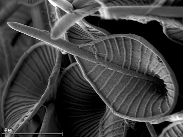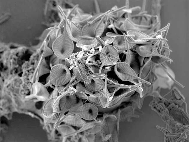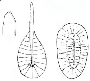
Pterocystis striata (Nicholls, 1983) Siemensma and Roijackers, 1988
Diagnosis. Cells 7-10 µm in diameter (fixed), with two kinds of scales. Spine-scales a hollow shaft, 3-7 µm long, 0.12-0.2 µm wide and tapered bluntly to a rounded apex. The proximal half of the spine shaft is bordered by an obovoid membrane with a peripherally thickened margin about 0.02 µm wide. The surface of the membrane is traversed with 20-42 radiating ribs (0.02 µm wide) connecting the margin of the membrane with the proximal half of the spine shaft. The base of the spine shaft is bent, drawing the central area of the membrane into a depression and imparting a spoon- or ladle-shaped appearance to the spine-scale. Plate-scales elliptical (1.4 x 2.2 µm) with a median tubular thickening approx. 0.07 µm wide with tapered ends and two-thirds the length of the scale. Edge of the scale ordered by a broad fold or thickened margin ca. one half the width of the scale and patterned with numerous radially disposed ribs 0.02 µm wide. Similar ribs connect the median thickening to the scale margin.
Remarks: This species can be confused with A. pteracantha.