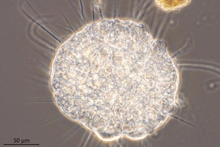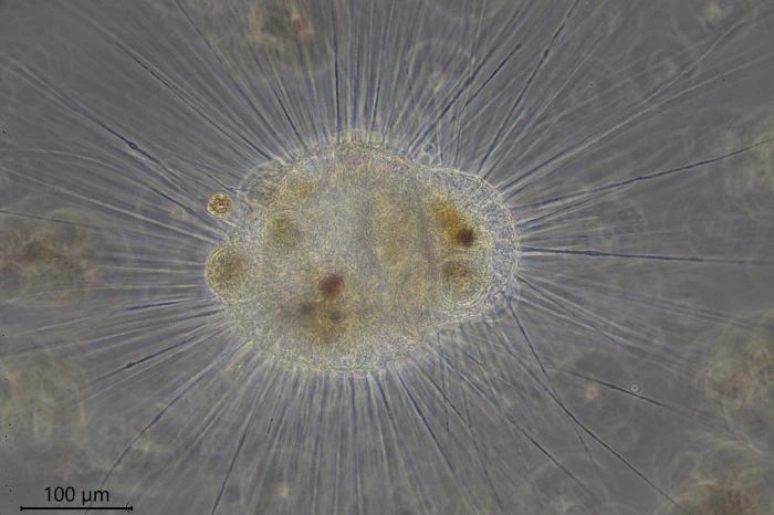
Actinosphaerium nucleofilum Barrett, 1958
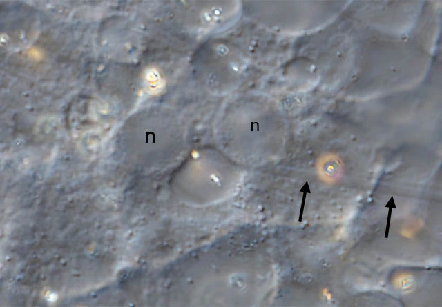
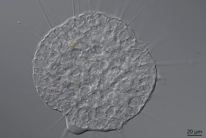
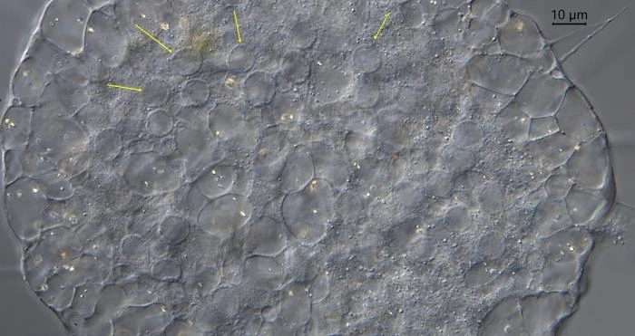
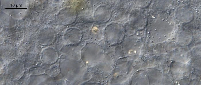
Diagnosis: Cell strongly vacuolated, ecto- and endoplasm not clearly differentiated. Diameter of endoplasm approximately the thickness of the ectoplasm. With small and numerous nuclei, 4-11 µm in diameter, with peripherally located nucleoli and with a cyst wall incorporating spherical siliceous elements. Axopodia approximately twice as long as the cell diameter. Cells can adhere so strongly to the substrate that they cannot be aspirated with a micro pipette!
Dimensions: Cell body 45-700 µm, average 200-400 µm. My measurements 230-400 µm; nuclei 4-11 µm.
Ecology: Freshwater. North-America (Barrett, 1958) and Europe (Netherlands, Siemensma, unpublished, 1981 and 2020; Corsica, Siemensma, unpublished, 2022)
Remarks: Barrett (1958) distinguished this species from A. eichhornii, because in A. nucleofilum the axopodia terminate on the surface of a nucleus or in their immediate vicinity, and the nucleolar material is located peripherally. The diameter of the nuclei is relatively small. Cysts have spherical siliceous elements in the wall (Patterson and Thompson 1981).