Cylindrifflugia lanceolata
C. lanceolata, shells 153-169 µm long, nuclei 23-26 µm (Naardermeer, Netherlands)

Cylindrifflugia lanceolata (Penard, 1890)
Basionym: Difflugia lanceolata Penard, 1890

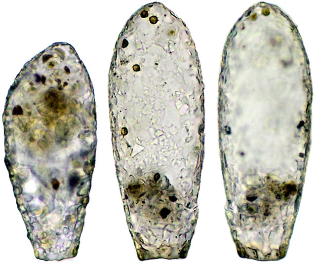
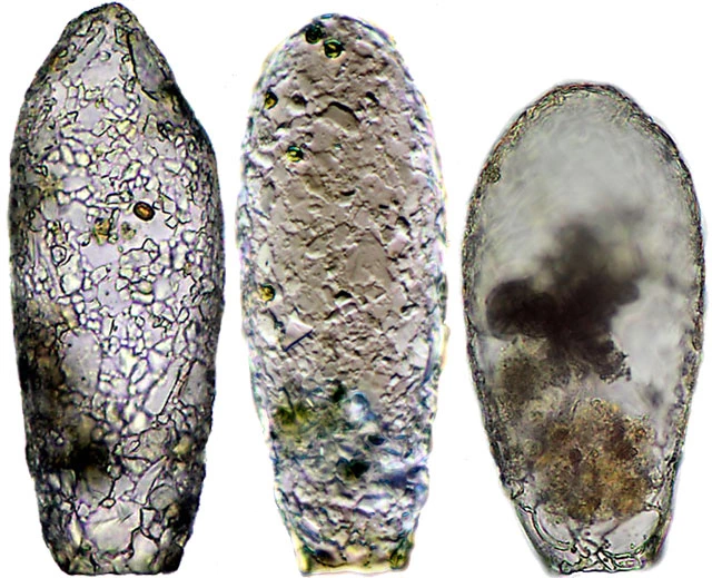
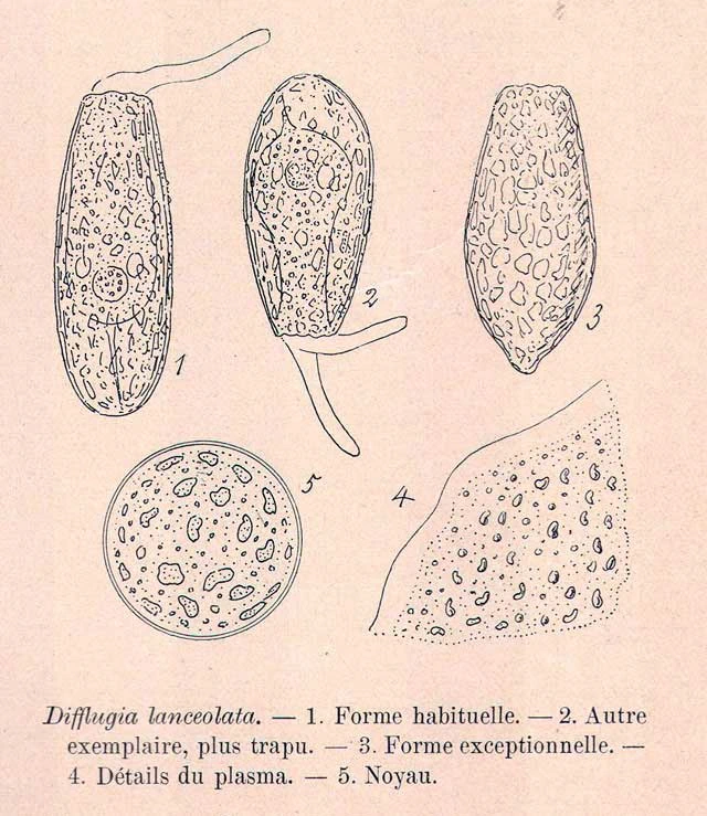
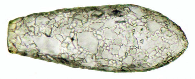
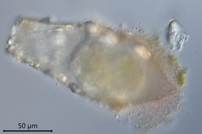
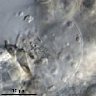
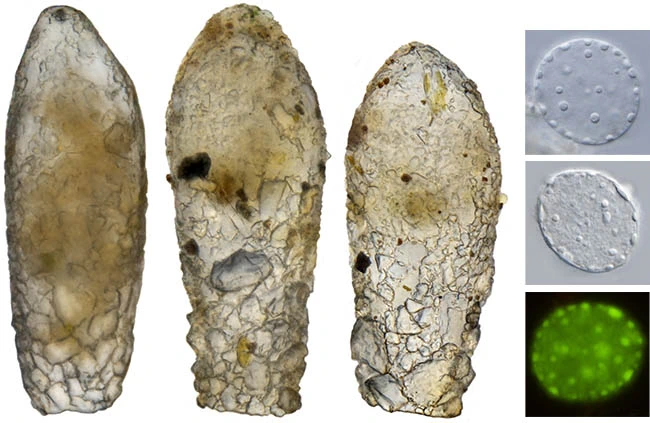
Diagnosis: The shell is lanceolate, not compressed, and displays a highly regular contour. It is hyaline, thin, and chitinoid, consistently covered with flat, thin siliceous particles that give the surface a polished and smooth appearance. The fundus is typically rounded, though it may occasionally exhibit a more or less pronounced ogival shape or, in rare instances, form a terminal nipple. The aperture is circular and often bordered by an organic collar. The nucleus is ovular, containing small, roundish nucleoli, some of which are flattened against the nucleolar membrane. Zoochlorellae are absent.

Dimensions: Penard (1902): Shell length 140—160 µm; nucleus ≈28 µm. My measurements: 138—200 µm; nucleus 23—26 µm (n=3).

Ecology and distribution. Freshwater, several water types. I found it also in eutrophic water, in ditches between heavily fertilized farmland. Chardez (1978) found that “D. lanceolata and D. penardi are species which are sometimes found in limnosaprobic muds (…)”.

Remarks: Shells of this species are easily recognizable because of their smooth surface, though also specimens with rougher xenosomes are found. Sometimes the fundus is a little asymmetrically pointed, but the clean outline of this species is the main distinguishing feature,

Cylindrifflugia lanceolata
Shells 143-192 µm. All shells were found in a sample from a pond in the Odenwald, Germany, 2024.
Cylindrifflugia lanceolata
C. lanceolata, nuclei, three in DIC, one in Fluorescence (stained with Acridine Orange)
Cylindrifflugia lanceolata
Shells 145-170 µm long, all from the same sample, Den Treek, Amersfoort – Photos stacked
Cylindrifflugia lanceolata
Shells 154-174 µm long; the first two shells from Den Treek, the right one from the Laegieskamp. The second shell was photographed in DIC.
Cylindrifflugia lanceolata
Shells resp. 200, 185 and 130 µm. All three shells were found in a sample from a location near the city of Lemmer, Friesland.
Cylindrifflugia lanceolata
Drawing: Penard, in Faune Rhizopodique du Bassin du Léman, 1902
Cylindrifflugia lanceolata
C. lanceolata, slide 262-2-3 from the Pénard Collection in Geneva – Photographed by Thierry Arnet, as part of the Pénard project (https://creativecommons.org/licenses/by-sa/3.0/deed.en).
Cylindrifflugia lanceolata
C. lanceolata, slide 260-9-4 from the Pénard Collection in Geneva – Photographed by Thierry Arnet, as part of the Pénard project (https://creativecommons.org/licenses/by-sa/3.0/deed.en).
Cylindrifflugia lanceolata
C. lanceolata, slide 261-9-1 from the Pénard Collection in Geneva – Photographed by Thierry Arnet, as part of the Pénard project (https://creativecommons.org/licenses/by-sa/3.0/deed.en).
Cylindrifflugia lanceolata
C. lanceolata, 156 µm
Cylindrifflugia lanceolata
C. lanceolata, with parasite; nucleus 22 µm.
Cylindrifflugia lanceolata
C. lanceolata, shell 190 µm, nucleus 28 µm.
Cylindrifflugia lanceolata
C. lanceolata, with distinct organic collar – 138 µm
Ferry Siemensma, created July 3, 2022; last modified February 18, 2026
Recent posts