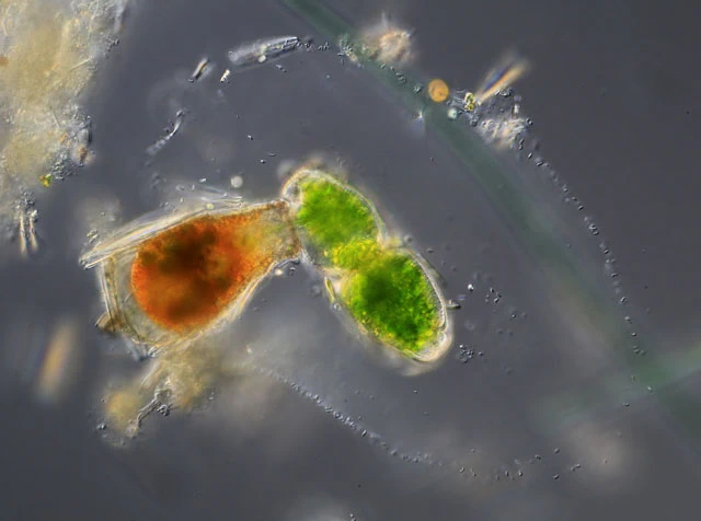
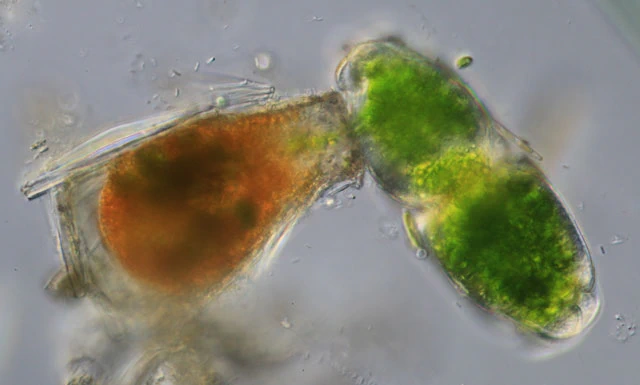
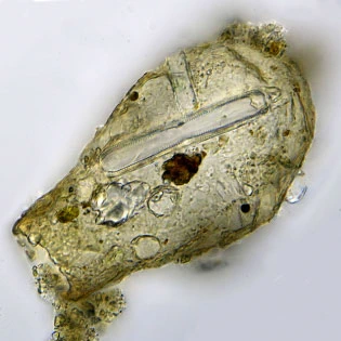
Pseudonebela africana Gauthier-Lièvre, 1953
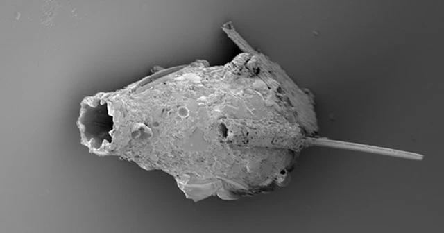
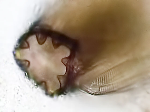
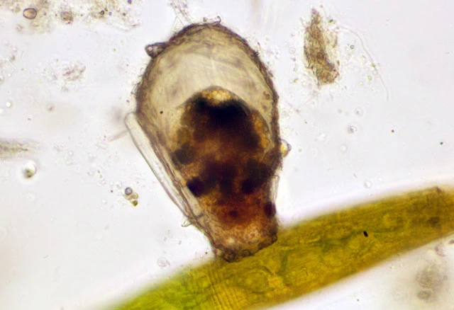
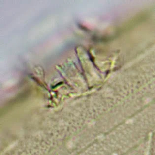
Diagnosis: Shell lageniform and circular in cross-section; dorsal region rounded or slightly oval, composed of organic secretion of an apparently smooth cement and agglutination of extraneous particles including diatoms and, more commonly, many rounded plates, and with many small irregular platelets juxtaposed in between the larger rounded plates, thus resembling a Nebela-species, hence the name Pseudonebela; it’s unclear whether the plates are obtained from the environment or produced by the amoeba; aperture very characteristic by an organic covering with 4-11 small inward curving lips which appear as ‘denticles’ under optical microscopy, creating a characteristic cross- or clover-shaped aperture; electron microscopy reveals that these ‘denticles’ are not minute plates as originally described (Gauthier-Lièvre, 1953), but rather are invaginations of the aperture’s edge. Nucleus single, large, with fragmented nucleolar material, concentrated in some irregularly shaped pieces of variable size.
Dimensions: Shell 90–100 µm long, 50–60 µm wide, aperture 25–28 µm (Gauthier‑Lièvre, 1953); approximately 87 µm long, 46 µm wide, aperture 27 µm (Lahr & Gomes e Souza, 2011); 85–185 µm long, nucleus 29–35 µm (Siemensma & Opitz, 2014).
Ecology: Freshwater; shallow ponds with aquatic vegetation, as well as swamps and Sphagnum vegetation.
Distribution: Reported from Africa (Burkina Faso and Angola, 1953), South America (five localities in central and southeastern Brazil, 2011), Florida (2012), Austria (2013, A. Opitz, pers. comm.), and China (Siemensma, unpubl. 2015).
Remarks: Pseudonebela africana is considered a flagship species—its morphology is so distinctive that it cannot be confused with any other known species. It was long thought to have a restricted geographic distribution. As noted by Lahr and Gomes e Souza, the characteristic aperture may have been overlooked in earlier studies, as careful examination is required to identify P. africana with confidence. It may be confused with other lageniform species such as Nebela lageniformis, Difflugia bacillifera, and Difflugia rubescens. The species has recently been recorded from the Northern Hemisphere (Florida, USA, and Austria, Europe). See Siemensma & Opitz (2014).


Original description:
Thèque lagéniforme non comprimée dont l’aspect général est celui de la Nebela Americana Taranek. Panse largement arrondie ou ovoïde surmontée d’un col trapu généralement cylindrique, plus ou moins long, terminé par un pseudostome tronqué droit. Ce pseudostome est bordé d’un bourrelet chitininoïde épaissi débordant sur l’ouverture qu’il rétrécit assez fortement, il porte 3-5 petites dents formées chacune d’une minuscule plaquette triangulaire; l’ouverture apparait 3-5 lobée.
Sauf le bourrelet du pseudostome qui est plus ou moins jaunâtre et légèrement granuleux, la thèque est incolore et d’une extrême transparence. Le revêtement consiste généralement en plaquettes circulaires ou ovales, rarement juxtaposées, mais empiétant le plus souvent largement les unes sur les autres; la présence de plaquettes de renforcement plus petites est fréquente. Les plaquettes de recouvrement peuvent également être polygonales irrégulières, juxtaposes et unies par un ciment chitinoïde souvent dispose en ponts donnant un aspect ponctué au contour des plaquettes.
Dimensions: 90-100 X 50-60 µm; pseudostome 25-28 µm; hauteur du col 30 µm.
Haute-Volta, lac de Banfora, très commune dans la marge d’hélophytes. Aussi trouvé dans une récolte du Sénégal.