Pelomyxa gruberi Frolov, Goodkov, Chystjakova and Skarlata, 2006
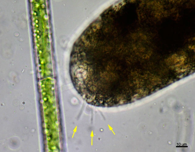
Diagnosis: Rounded (feeding stages) or elongated cylindrical (locomotive stages) amoeboid organisms. A well-developed broad zone of hyaline cytoplasm with slender finger-like hyaline protrusions formed at the anterior body end during locomotion. Cell diameter varies from 80 µm (young uninucleate individuals) to 350 µm (multinucleate locomotive forms). In the life cycle, uninucleate developmental stages alternate with multinucleate ones. Reproduction occurs by plasmatomy of multinucleate amoebae: they form division rosettes or divide unequally. The cytoplasm clearly differentiated into ectoplasm and endoplasm. Vesicular nuclei (1-32 per cell) with a single central nucleolus. Nuclear envelope surrounded with microtubules in all life cycle stages. Flagella are numerous, non-motile; axoneme with a non-standard microtubular pattern. In the cytoplasm two types of rod-shaped endocytobionts are present: (1) large bacteria with a pronounced longitudinal cleft, and (2) smaller methanogen-like bacteria. With a light microscope considerable agglomerations of rodlike bacteria radiating from the nuclear surface into the cytoplasm can be seen. No glycogen bodies are formed in any life cycle stages. Cytoplasm color varies from light beige to dark brown.
Dimensions: The size of uninucleate P. gruberi individuals varies from 79.2 to 215.1 µm. These are spherical or slightly oval in shape. Nuclei: 19.7 to 52.1 µm, size depending upon the size of the cell and the number of nuclei present. My measurements: locomotive forms 160-240 µm, nuclei 25-29 µm.
Habitat: Amoebae feed on small detritus particles, mostly of vegetative origin. Pelomyxa gruberi can be found in water bodies of the North-West Russia throughout the year. These amoebae are silt dwellers at a depth of 5-70. They are always surrounded with detritus particles, adhering to short hyaline pseudopodia. P. gruberi feeds on small detritus particle of vegetative origin. Mineral inclusions in the cytoplasm are infrequent. The cytoplasm is light beige to dark brown, depending on the prevailing food inclusions, which makes the amoebae difficult to distinguish inside silt lumps or during cursory examination of samples.
I found this species in the Diepveen, together with Pelomyxa palustris.