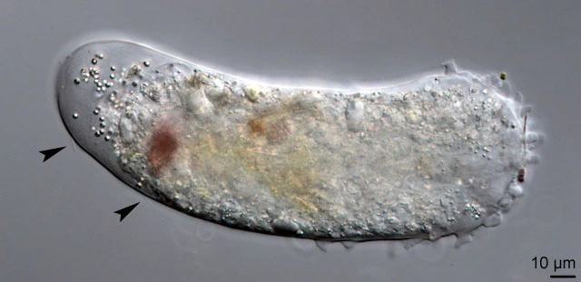
Pelomyxa spec., c. 206 µm long, with arrowed cilia and some glycogen bodies
Pelomyxa spec.
I found one specimen in July 2014 in the ooze of a very shallow pond. There where about 15 spherical nuclei, 9.8-13.4 µm in diameter, with a number of nucleoli in the periphery. The anterior end of the cell was a broad hyaline zone. There was no distinct fountain streaming as typical for Pelomyxa species. The anterior part showed some conical hyaline subpseudopodia. Some immobile cilia were present.

Some nuclei are visible