Amphifila marina (Dykstra & Porter, 1988)
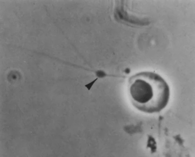
Diagnosis: Cells ovoid, colorless, containing one to several prominent eccentric refractive granules, a single uninucleolate nucleus, and one to several vacuoles. Cells moving with an irregular gliding motility by means of fine filamentous, branched ectoplasmic elements extending up to 35 µm from the polar extremities of the cell. Cell wall composed of overlapping Golgi-derived scales 1.5-1.9 µm in diam. Repeated cell divisions produce flat circular patches of cells in culture. Sporangia, spores and cysts not observed.
Dimensions: Cells 3.7- 5.9 x 5.1 – 8.5 µm.
Ecoloy. Known from western and eastern coastal waters of the United States. Emergent onto agar media from marine algae and vascular plant fragments. Associated with marine vegetation, most likely as a superficial saprotroph.
Remarks: Amphifila marina cells resemble very closely the vegetative cells of Sorodiplophrys stercorea (see micrograph below). Both organisms contain prominent refractive granules, uninucleolate nuclei, and finely branched ectoplasmic elements extending from the poles of the cells and possessing occasional swellings.
Taxonomic status unclear.
