D. biconcava, 157 µm long, in front view and lateral view –
Naardermeer Difflugia biconcava Ertl, 1965
Diagnosis: Shell ovoid or pyriform, with an aboral rounded cone or spine and laterally compressed. In lateral view, the shell exhibits a typical biconcave shape. The aperture is circular. The shell is composed of a mixture of small to large pieces of primarily flattish quartz, arranged to form a relatively smooth surface. The nucleus is spherical with a large central nucleolus containing multiple lacunae.
Dimensions: Ertl (1965): Shell length 100-143 µm; width 66-96 µm; aperture 29-32 µm. My measurements: Naardermeer 149-255 µm (n=10); nucleus 31-39 µm; Montfoort 78-104 µm. This species is extremely variable.
Habitat: Ertl found the type material in rice field samples from Slovakia. I encountered this species in sediments of a ditch near the Naardermeer, the Netherlands.
Remarks: D. balcanica Ogden and Zivkovich, 1983 is a very similar, but much smaller species. Ogden gives as length 111-114 µm and as width 79-82 µm (n=2).
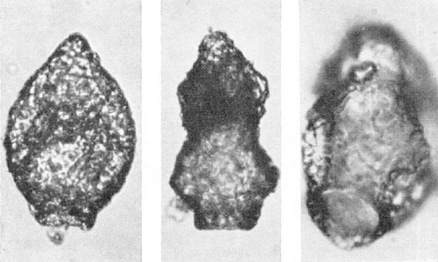
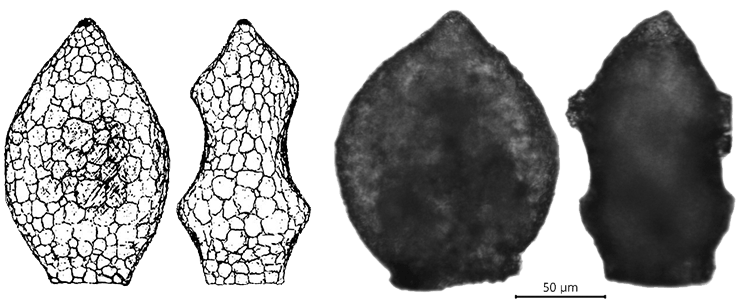
Difflugia biconcava, in frontal and lateral view – after Ertl, 1965 (left) and from
Naardermeer, 2015 (right).
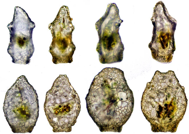
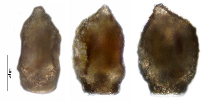
Difflugia biconcava, in lateral view (upper row) and frontal view (lower row. All shells came from the same sample and were alive. Notice the right one with two cones. Length 161-185 µm.
Difflugia biconcava, published by Ertl, 1965
D. balcanica, published by Ogden and Zivkovic, 1983. This species is very similar to D. biconcava, but much smaller: 111-114 µm.
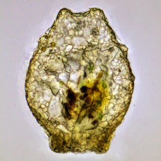
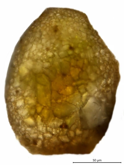
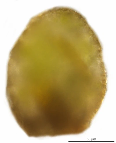
Difflugia biconcava, stacked image, shell 190 µm, Naardermeer
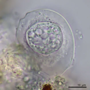
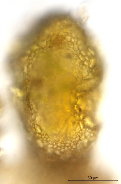
Difflugia biconcava, nucleus and pseudostome (Naardermeer)
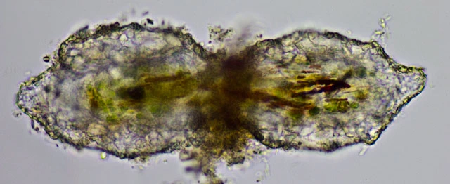
Two dividing cells, note the difference in size between them.
Difflugia biconcava, the same test in frontal and in lateral view.
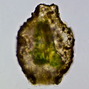
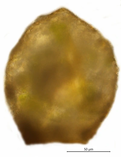
Difflugia biconcava
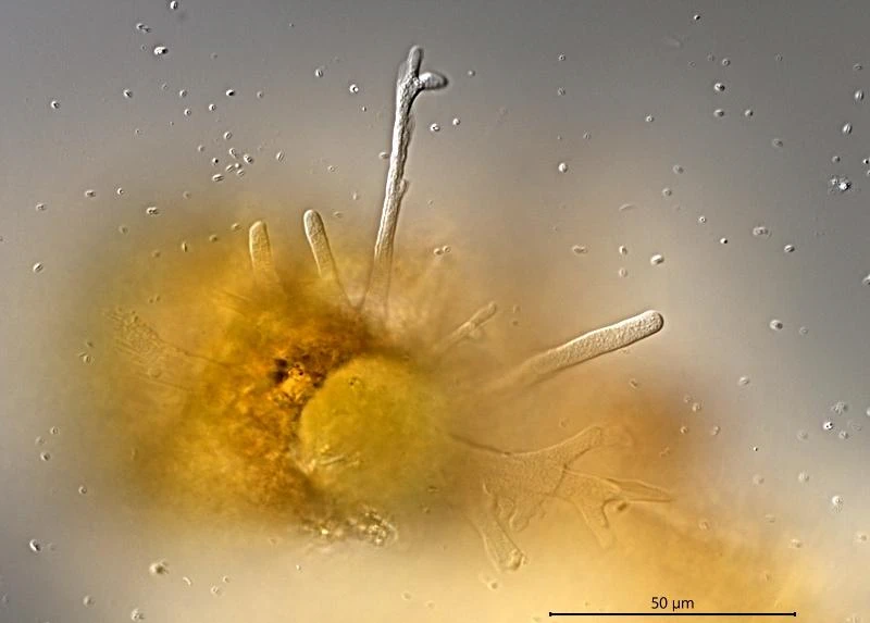
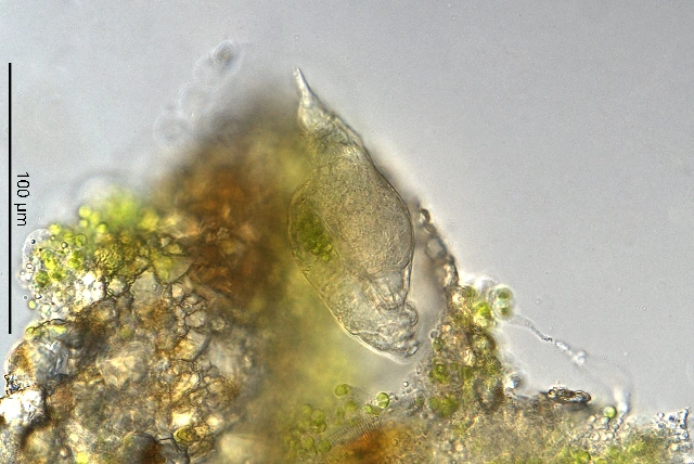
Difflugia biconcava, pseudopodia
Difflugia biconcava, in frontal and lateral view – after Ertl, 1965 (left) and from
Naardermeer, 2019 (right).
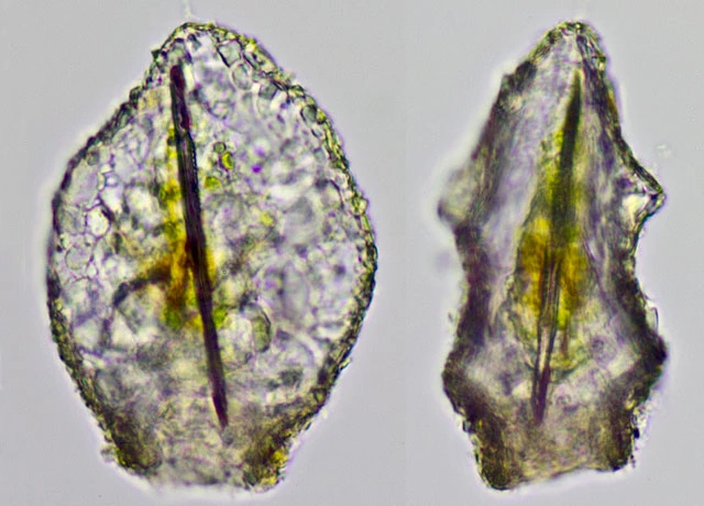
Difflugia biconcava, in lateral and frontal view, from
Naardermeer, 2025.
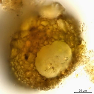
When a shell was crushed in a wet mount, a parasitic rotifer became visible, attempting to escape from the fragments (
Naardermeer, 2025)