Difflugia lobostoma parasitism
While attempting to identify several Difflugia specimens, I broke their shells by gently pressing the cover glass with a needle. With a bit of luck, the nucleus slips out of the test and can be observed. I use this as a key character to distinguish between D. gramen and D. lobostoma. When I broke the shell of what appeared to be D. lobostoma—based on the structure of the nucleus—a rotifer emerged from the test together with a rotifer egg.
References:
De Smet, W.H., 2006. Asciaporrectidae, a new family of Rotifera (Monogononta: Ploima) with description of Asciaporrecta arcellicola gen. et sp. nov. and A. difflugicola gen. et sp. nov. inhabiting shells of testate amoebae (Protozoa). Zootaxa 1339: 31–49 (2006)
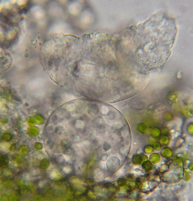
This is the young rotifer (above), together with the nucleus (below) and the zoöchlorellae that slipped out of the broken test. The test was 161 µm long; the egg (not visible here) was oval and 49 µm long. The rotifer, when fully stretched, measured 129 µm, and the nucleus 44 µm.
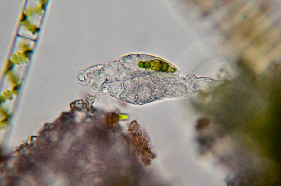
A second D. lobostoma test also contained a living amoeba and a young rotifer. This rotifer had zoöchlorellae in its stomach. This phenomenon reminded me of some older drawings I made in the 1970s. In that case, I had observed several specimens of D. capreolata containing many rotifer eggs as well as young rotifers. I think the rotifers hatch from the eggs and feed on part of the plasmal body. The rotifer in D. lobostoma resembles the one I observed in 1977 in D. capreolata.

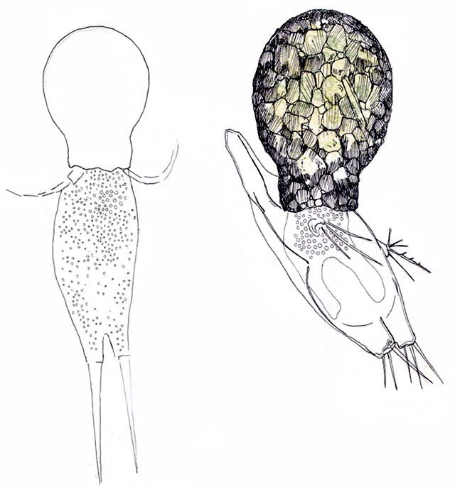
Difflugia capreolata – specimen upper- left with three rotifer eggs in its shell (1977).

A Difflugia capreolata:
A — test containing 13 eggs and two young rotifers
B — deformed nucleus of D. capreolata
C — rotifer that emerged from the test
D — rotifer egg
E — young rotifer with zoöchlorellae
F — chewing apparatus (mastax) of the rotifer
I have observed the following: a. An active specimen of D. capreolata with, inside the test (between body and fundus), four eggs and a young rotifer. Eggs approx. 70 µm long; rotifer approx. 160 µm. b. An empty D. capreolata test filled with 13 rotifers (Proales sp.?), one large specimen and 12 small ones. The large rotifer was approx. 210 µm long. c. A test with three eggs and two active rotifers. The test also contained a piece of protoplasm with a nucleus. The plasma formed pseudopodia. d. A test with one rotifer and four eggs, including protoplasm. e. A test with two rotifers and 13 eggs (see drawing A above), and protoplasm with zoöchlorellae. Both rotifers had zoöchlorellae in their stomachs.
I also observed that D. capreolata sucks in rotifers. On one occasion I saw a specimen empty a rotifer in ten minutes.