
P. fascicularis, after Penard, 1902
Pseudodifflugia fascicularis Penard, 1902
Diagnosis: shell chitinoid, pyroid, narrow elliptical, with a faint indication of a neck, not compressed, partly covered with some mineral particles and diatom frustules; Plasmaboy visible through the shell wall. Pseudosome circular, but hard to observe because of a bundle (‘fascis’) of mineral particles; cytoplasm and nucleus normal; filopodia long and straight, branching but not really anastomosing.
Dimensions: literature 17-50 µm, seldom longer. My measurements: 24 µm.
Ecology: Detritus.
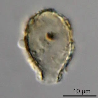

Pseudodifflugia fascicularis, shells 21 and 24 µm large

Pseudodifflugia fascicularis, filopodia

From: Lueftenegger et al, 1988