Centropyxis spinosa
Centropyxis spinosa, with 15 spines, Killarney National Park, Ireland, 2026

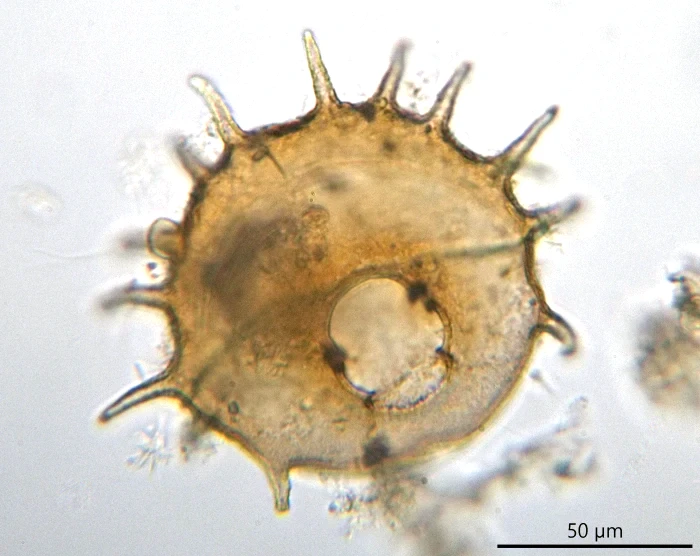
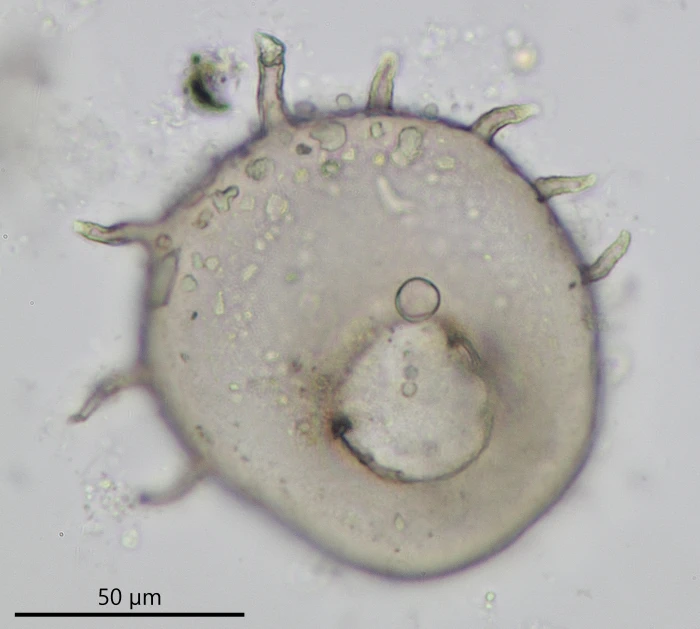
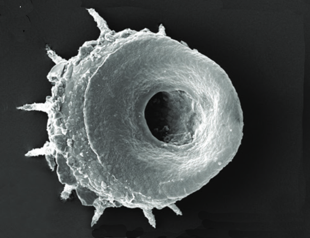
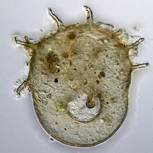
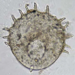
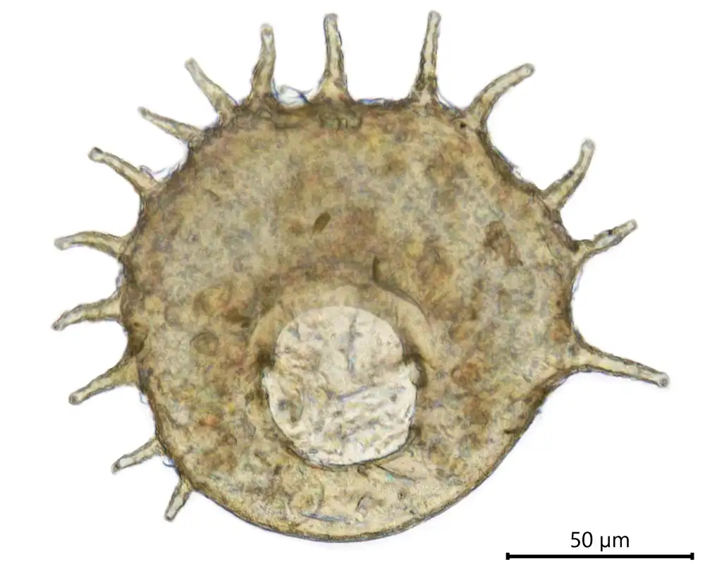
Centropyxis spinosa  (Cash, 1905)
Basionym: Centropyxis aculeata var. spinosa Cash, 1905
Synonym: Centropyxis bryophilus Dekhtyar, 1998

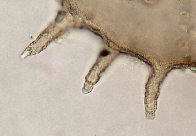
Diagnosis: Shell oval, with a neatly rounded anterior outline, purely chitinous, usually bearing relatively few adhering sand grains, and semitransparent; yellowish‑brown when young, gradually darkening with age, and sometimes partly or entirely covered with diatom frustules. Aperture usually oval, with the long axis parallel to the long axis of the shell, lobed or uneven in outline, variable in width, but always comparatively smaller than that of C. aculeata. The margin is reversed and usually has two bridges connecting the invaginated aperture with the opposite dorsal area. Spines are variable in number and length, composed of the same material as the test, and often curved, with a plug in the aperture at the upper end. A broad ventral region around the aperture has a characteristic smooth and uniform structure that is clearly visible under SEM and light microscopy.

Dimensions: Cash (1905): 120–140 µm.
Todorov and Bankov (2019): length 107–136 µm; height 42–46 µm; spines 14–28 µm; aperture diameter 28–37 µm.
My measurements: length 91–117 µm; L/W ratio 0.9–1.2; aperture 25–45 µm × 21–36 µm.

Ecology: In Sphagnum, or in water bodies in contact with Sphagnum.

Remarks: The lobed aperture is caused by the presence of two pillar‑like internal bridges and an anterior crescent‑shaped connection between the ventral and dorsal regions, resembling a headphone (see images below).

Centropyxis spinosa
Centropyxis spinosa, all specimens from a single sample collected in Sphagnum at Oppad, Kortenhoef, Netherlands (2025)
Centropyxis spinosa
Centropyxis spinosa in lateral view, with inner structure, Oppad (2025)
Centropyxis spinosa
Centropyxis spinosa in lateral view, with inner structure, 99 µm, Oppad (2025)
Centropyxis spinosa
Centropyxis spinosa , with two struts clearly visible, Norway (2023)
Centropyxis spinosa
Centropyxis spinosa , showing a strongly lobed aperture
Centropyxis spinosa
Centropyxis spinosa , Tirol, Austria (2016)
Centropyxis spinosa
A broad ventral region around the aperture typically exhibits a smooth and uniform structure when observed under SEM (photomicrograph: Todorov and Bankov, 2019). This feature is also clearly visible under light microscopy (see photograph below), although not in all shells.
Centropyxis spinosa
C. spinosa , ventral view of the smooth surface
Centropyxis spinosa
Centropyxis spinosa, 97 µm, with small stones in the spines
Centropyxis spinosa
Centropyxis spinosa, 120 µm, with small stones in the spines
Centropyxis spinosa
Centropyxis spinosa
Centropyxis spinosa
Centropyxis spinosa
Centropyxis spinosa
Centropyxis spinosa
C. spinosa, left: after Cash, 1905; right: 101 µm
Centropyxis spinosa
Centropyxis spinosa
Centropyxis spinosa, 95 and 100 µm, exclusive spines
Centropyxis spinosa
Centropyxis spinosa
Centropyxis spinosa, both 120 µm, inclusive spines
Ferry Siemensma, created February 23, 2019; last modified February 21, 2026
Recent posts