Centropyxis todorovi Siemensma, Shimano & Wanner, 2025
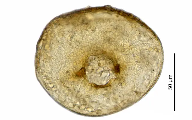
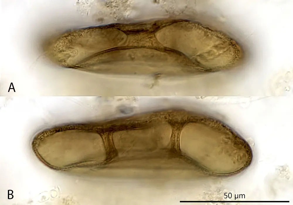
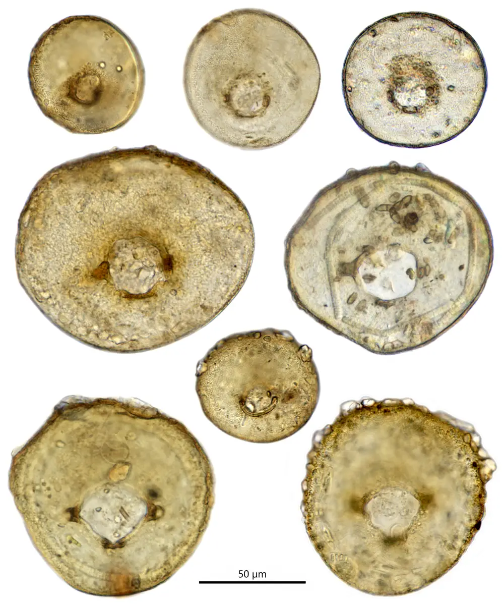
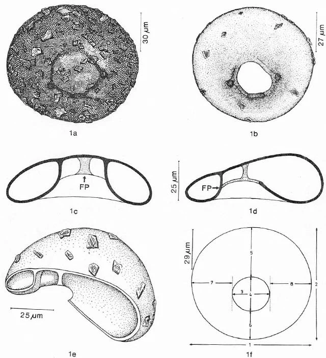
Diagnosis: Shell organic, yellow to brown or brownish-grey, and transparent. In ventral view, the shell is broadly oval to circular. In lateral view, the shell height is less than half the shell diameter and is strongly compressed towards the frontal area. The aperture is eccentrically located, invaginated, and usually equipped with three bridles or struts that connect the invagination to the ceiling of the shell. These bridles form the vertices of an isosceles triangle, with the apex pointing towards the frontal area of the shell. The base of this triangle roughly aligns with the midline of the aperture. The organic cement of the ventral area is smooth and lacks embedded xenosomes, whereas the dorsal area contains mineral particles, which increase in size towards the posterior part.
Dimensions: Shell diameter 60-112 µm.
Habitat: Sediment of ditches, among aquatic plants, and in Sphagnum.
Geographical distribution: Netherlands (Siemensma, pers. observations), Japan (Wanner et al., 2025); Germany (Schönborn et al., 1983; Odenwald, Siemensma, 2024); Austria (Schönborn et al., 1983); India (Chattopadhyay and Das, 2003; Farooqui et al., 2020; Purushothaman and Bindu, 2015); Bulgaria (Todorov and Bankov, 2019).
Differential diagnosis: Centropyxis todorovi can be distinguished from all known bridged Centropyxis species by the presence of three internal bridges arranged in the form of a triangle with the apex pointing forward.
Remarks: This species is fairly easy to identify due to its three supporting pillars. Both small and large forms were found in the same sample from a pond in the Odenwald, Germany. The large shells resemble those illustrated and described by Schönborn et al. (1989)—see the bottom of this page—but were erroneously identified as C. laevigata. Chattopadhyay and Das (2003), Todorov and Bankov (2019), and Farooqui et al. (2020) also identified their specimens as C. laevigata, while Purushothaman and Bindu (2015) attributed theirs to C. delicatula Penard, 1902.
However, C. laevigata Penard, 1890, differs significantly from the shells described by these authors. Penard (1890) described a high dome that is barely compressed anteriorly (“un dôme assez élevé et à peine comprimé en avant”), with measurements of 120–150 µm in diameter and 80–120 µm in height. The ventral area features a deep, inward-invaginated, eccentrically located round aperture, connected to the opposite dorsal wall by a single broad chitinous band covered with thick, amorphous scales (“une large bride foncée couverte d’écailles morphes épaisses”). Penard’s drawings clearly illustrate these features (Penard, 1890, 1902). This single band or bridge is a defining characteristic of the genus Frenopyxis Bobrov & Mazei, 2020, and consequently, Wanner et al. (2025) transferred C. laevigata to that genus.
Centropyxis delicatula Penard, 1902, is another species with internal bridges. Penard (1902) described it as very small, with a diameter of 35–48 µm. This species, characterized by a predominantly organic shell, is relatively flat and has an inward-facing construction around the aperture, featuring four to five bridges connecting the ventral side to the opposite inner dorsal wall. At the posterior side of the aperture, adjacent to the fundus, there are always two and sometimes three bridges, while at the anterior side, there are consistently two shorter bridges. The posterior bridges converge at the top, forming a rounded window. Occasionally, Penard (1902) observed one or two spines at the rear of the shell. The internal framework resembles that of Centropyxis lapponica (Łuców et al., 2025, Fig. 7f.). Micrographs by Purushothaman and Bindu (2015), attributed to C. delicatula, show three bridges arranged in a triangular position. These shells do not match the original description of C. delicatula but instead resemble C. todorovi.
It is likely that C. laevigata and C. delicatula have been misattributed to other species in the past. Tripathi et al. (2017) published a micrograph of a shell with bridges and spines labeled as C. laevigata (their Fig. 8h). However, the presence of spines and the number of bridges fall outside the diagnosis of C. laevigata. Similarly, their Fig. 8g depicts a shell with four bridges. Chattopadhyay & Das (2003) reported C. laevigata and noted the presence of bridges but provided drawings without any indication of these structures, making it difficult to assess their observations. The same issue applies to the drawings published by Golemansky (1966) as C. laevigata.

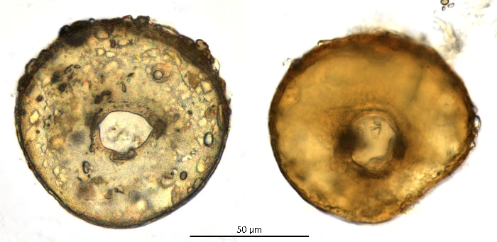
The species below was described by Schönborn et al, 1983, as C. laevigata, but is clearly a different species, as C. laevigata is clearly defined by the presence of one single broad ribbon or bridge, which places that species in genus Frenopyxis.
Schönborn et al. (1983) gave the following diagnosis:
Shell ellipsoid, rarely almost circular, flattened to a ratio of 2-3:1, slightly more pronounced in the frontal region than in the rear, resulting in a wedge-shaped side view. In a section through the longer axis, however, the shell is evenly curved. Pseudostome regularly to irregularly ellipsoid, always clearly a-centrally positioned, resulting in a narrower frontal and a wider rear shell section. On the longer shell axis, however, it lies on average in the center of the shell axis: ventral side in the frontal region towards the mouth slopes very steeply dorsally, but in the rear region it tapers off moderately steeply. Due to the steep slope in the frontal region, the pseudostome edge appears thickened when viewed from above. The ventral shell wall forms a wider frontal pillar and two narrower lateral pillars at the aperture, which are attached to the inner wall of the dorsal side. In dorsal and lateral views, the shell is more or less clearly pitted at the attachment points of the three pillars.
Young shells are light brown, older ones dark brown, sparsely to densely covered with thin, colorless xenosomes, presumably quartz plates, usually somewhat less densely ventrally than dorsally. The plates are often so thin that the underlying shell structure can still be seen using both light and scanning electron microscopy. The shell is quite soft, flexible, with hexagonal to polygonal fields. Fields approximately 0.5 µm in size, also easily visible under light microscopy at high magnification.
References:
Schönborn, W., Foissner, W. and Meisterfeld, R. (1983): Licht- und Rasterelektronenmikroskopische Untersuchungen zur Schalenmorphologie und Rassenbildung Bodenbewohnender Shellaceen (Protozoa : Rhizopoda) sowie Vorschläge Zur Biometrischen Charakterisierung von Shellaceen-Schalen – Protistologica, XIX, 4, p. 553-566.